 뒤로
Skeletal Muscle Tissue and the Muscular System: Structure, Function, and Physiology
스터디 가이드 - 스마트 노트
 자료에 맞춘 맞춤형 노트, 핵심 정의, 예시, 맥락을 확장해 제공합니다.
Skeletal Muscle Tissue
Types of Muscle Tissue
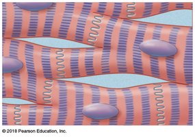
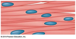
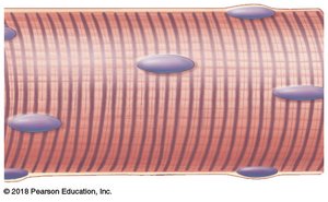
There are three main types of muscle tissue in the human body, each with distinct structural and functional characteristics:
Cardiac Muscle: Involuntary, found only in the heart, branched and striated with intercalated discs for synchronized contraction.
Smooth Muscle: Involuntary, located in the walls of hollow organs and blood vessels, spindle-shaped with a single nucleus, non-striated.
Skeletal Muscle: Voluntary, attached to bones, composed of long cylindrical fibers with multiple nuclei and visible striations. Responsible for movement by acting on the skeleton as levers.
Functions of Skeletal Muscle
Produce skeletal movement
Maintain posture and body position
Support soft tissues
Guard entrances and exits (e.g., sphincters)
Maintain body temperature (thermogenesis)
Store nutrient reserves (glycogen)
Structure of Skeletal Muscle
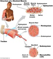
Skeletal muscle is a complex organ composed of muscle tissue, connective tissue, nerves, and blood vessels. The organization is hierarchical:
Epimysium: Dense collagenous layer surrounding the entire muscle.
Perimysium: Divides muscle into fascicles (bundles of fibers), contains blood vessels and nerves.
Endomysium: Surrounds individual muscle fibers, contains capillaries, nerve fibers, and myosatellite cells (stem cells for repair).
Development of Skeletal Muscle
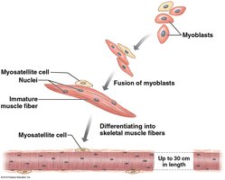
Skeletal muscle fibers develop from embryonic cells called myoblasts:
Myoblasts fuse to form large, multinucleate muscle fibers.
Some myoblasts remain as myosatellite cells in the endomysium, aiding in muscle repair and regeneration.
Mature muscle fibers can be up to 30 cm (12 inches) long and contain many nuclei, allowing rapid protein synthesis for contraction.
Muscle Fiber Structure
Each muscle fiber contains specialized structures for contraction:
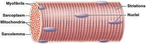
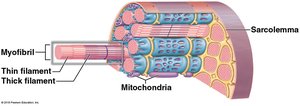
Sarcolemma: The cell membrane of a muscle fiber.
Sarcoplasm: The cytoplasm of the muscle fiber, containing organelles and myofibrils.
Myofibrils: Bundles of protein filaments (myofilaments) responsible for contraction.
Mitochondria: Provide ATP for contraction.
Striations: Alternating light and dark bands due to the arrangement of myofilaments.
Key Terms in Muscle Cell Structure
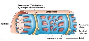
T-tubules (Transverse tubules): Invaginations of the sarcolemma that conduct action potentials deep into the muscle fiber.
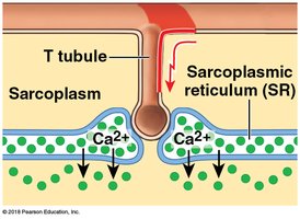
Sarcoplasmic Reticulum (SR): Specialized smooth endoplasmic reticulum that stores and releases calcium ions (Ca2+).
Myofilaments: Thin (actin) and thick (myosin) protein filaments organized into myofibrils.
Sarcomere Structure
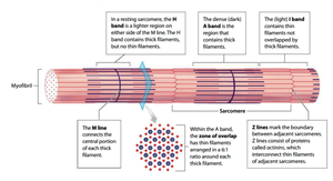
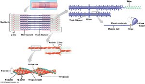
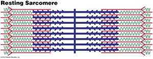
The sarcomere is the basic contractile unit of skeletal muscle, defined by the region between two Z-lines. The arrangement of actin (thin) and myosin (thick) filaments gives rise to the striated appearance of skeletal muscle.
Actin: Thin filament, anchored to the Z-line.
Myosin: Thick filament, anchored at the M-line.
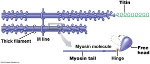
Titin: Elastic protein connecting Z-line to M-line, helps recoil sarcomere after stretching.
Thick and Thin Filament Structure
Thick Filament (Myosin): Composed of ~300 myosin molecules, each with a tail and two heads. The core contains titin for elasticity.
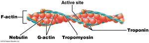
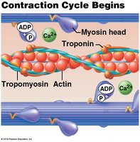
Thin Filament (Actin): Composed of F-actin (two strands of G-actin), nebulin (holds F-actin together), tropomyosin (covers myosin binding sites), and troponin (binds Ca2+ and moves tropomyosin).
Arrangement of Filaments in the Sarcomere
Thick and thin filaments are precisely organized within the sarcomere, creating distinct bands:
A band: Dark region containing thick filaments (with some overlap of thin filaments).
I band: Light region containing only thin filaments.
H band: Central region of A band with only thick filaments.
M line: Center of the sarcomere, where thick filaments are anchored.
Z line: Boundary between adjacent sarcomeres, where thin filaments are anchored.
The Sliding Filament Theory
The sliding filament theory explains how muscles contract:
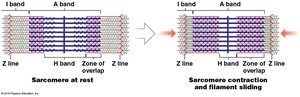
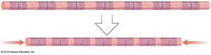
Thin filaments slide past thick filaments, shortening the sarcomere.
H and I bands decrease in width, while the A band remains constant.
Z lines move closer together, resulting in muscle contraction.
Muscle Cell Excitability and Contraction
Resting Membrane Potential
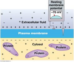
Muscle fibers maintain a resting membrane potential (typically -80 to -85 mV) due to the action of the Na+/K+ pump and the presence of large, negatively charged proteins inside the cell.
Na+/K+ pump: Moves 3 Na+ out and 2 K+ in, creating a polarized membrane.
Large proteins: Contribute to the negative charge inside the cell.
Action Potential and Depolarization
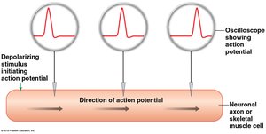
Muscle contraction is initiated by an action potential:
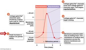
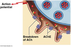
Ligand-gated Na+ channels: Open in response to acetylcholine (ACh) from a motor neuron, allowing Na+ influx and depolarization.
Voltage-gated Na+ and K+ channels: Open at threshold, causing rapid depolarization (Na+ in) and repolarization (K+ out).
Neuromuscular Junction (NMJ)
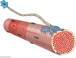
The NMJ is the synapse between a motor neuron and a skeletal muscle fiber:
Axon terminal: Releases ACh into the synaptic cleft.
Synaptic cleft: Space between neuron and muscle fiber.
Motor end plate: Region of sarcolemma with ACh receptors and Na+ channels.
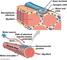
Sequence of Events at the NMJ
ACh is released from the motor neuron into the synaptic cleft.
ACh binds to receptors on the muscle fiber, opening Na+ channels and depolarizing the membrane.
ACh is broken down by acetylcholinesterase, ending the signal.
If threshold is reached, an action potential spreads across the sarcolemma and down T-tubules.
The sarcoplasmic reticulum releases Ca2+, initiating contraction (excitation-contraction coupling).
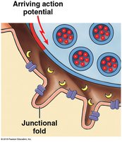
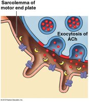
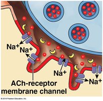
Excitation-Contraction Coupling
This process links the action potential in the sarcolemma to muscle contraction:
Action potential travels along sarcolemma and T-tubules.
Triggers Ca2+ release from the sarcoplasmic reticulum.
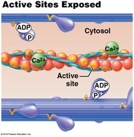
Ca2+ binds to troponin, shifting tropomyosin and exposing myosin binding sites on actin.
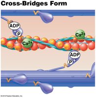
Myosin heads bind to actin, initiating the contraction cycle.
The Contraction Cycle
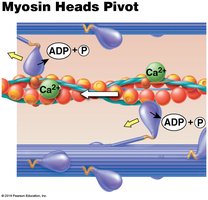
Myosin heads are energized by ATP hydrolysis (ADP and P remain attached).
Ca2+ binds to troponin, moving tropomyosin and exposing active sites on actin.
Myosin heads form cross-bridges with actin.
Myosin heads pivot, pulling actin filaments toward the M-line (power stroke), releasing ADP and P.
ATP binds to myosin, causing detachment from actin.
ATP hydrolysis re-cocks the myosin head, ready for another cycle.
Muscle Tension and Contraction Types
Muscle Tension
Muscle tension is the force generated during contraction. It depends on:
Number of cross-bridges formed (determined by sarcomere length and overlap of filaments).
Number of muscle fibers activated (motor unit recruitment).
Optimal overlap of thick and thin filaments produces the greatest tension.
Muscle Twitch and Phases
A muscle twitch is a single contraction-relaxation cycle in a muscle fiber, consisting of three phases:
Latent Period: Action potential triggers Ca2+ release.
Contraction Phase: Ca2+ binds troponin, cross-bridge cycling, tension rises.
Relaxation Phase: Ca2+ returns to SR, cross-bridges detach, tension falls.
Types of Muscle Contractions
Isotonic Contraction: Muscle changes length (shortens or lengthens) while tension remains constant.
Concentric: Muscle shortens as tension exceeds load.
Eccentric: Muscle lengthens as tension is less than load.
Isometric Contraction: Muscle length does not change; tension never exceeds load (e.g., postural muscles).
Energy for Muscle Contraction
ATP Sources
Free ATP: Immediate energy, enough for a few seconds of activity.
Creatine Phosphate: Transfers phosphate to ADP to regenerate ATP quickly.
Glycogen: Stored glucose, used in glycolysis and aerobic respiration for sustained ATP production.
Aerobic Cellular Respiration
Muscle fibers generate ATP through aerobic respiration when oxygen is available:
Glycolysis (cytoplasm): Glucose → pyruvate, small ATP yield, no O2 required.
Citric Acid Cycle (mitochondria): Pyruvate → CO2, NADH, FADH2.
Electron Transport Chain (mitochondria): NADH/FADH2 → ATP, requires O2.
Muscle Fatigue and Recovery
During peak activity, ATP demand exceeds supply; glycolysis predominates, producing lactic acid and leading to fatigue.
The Cori Cycle recycles lactate from muscles to the liver, where it is converted back to glucose.
Oxygen Debt: Extra oxygen required after exercise to restore normal conditions.
Muscle Fiber Types
Fast Fibers: Large, powerful, fatigue quickly, pale in color.
Slow Fibers: Smaller, slower to contract, resist fatigue, rich in myoglobin, red in color.
Intermediate Fibers: Characteristics between fast and slow fibers, more resistant to fatigue than fast fibers.
Clinical Correlations
Hypertrophy: Increase in muscle size due to repeated stimulation.
Atrophy: Decrease in muscle size due to disuse, aging, or nerve damage.
Muscular Dystrophy: Inherited diseases causing muscle weakness and degeneration (e.g., Duchenne, Becker).
Polio: Viral infection attacking motor neurons, causing paralysis.
Tetanus: Bacterial toxin causing sustained muscle contraction.
Botulism: Bacterial toxin blocking ACh release, causing paralysis.
Myasthenia Gravis: Autoimmune disease reducing ACh receptors, causing muscle weakness.
Rigor Mortis: Post-mortem muscle contraction due to ATP depletion and sustained Ca2+ presence.
The Muscular System
Organization of Muscles
Axial Muscles: Support and position the axial skeleton (head, neck, trunk).
Appendicular Muscles: Support, move, and brace the limbs.
Fascicle Organization & Leverage
Parallel Muscles: Fibers run parallel to the long axis (e.g., biceps brachii).
Convergent Muscles: Fibers converge on a common attachment (e.g., pectoralis major).
Pennate Muscles: Fibers attach at an angle to a central tendon (e.g., rectus femoris).
Circular Muscles: Fibers arranged in a circle (e.g., orbicularis oris).
Levers and Muscle Actions
First-Class Lever: Fulcrum between effort and load (e.g., neck extension).
Second-Class Lever: Load between fulcrum and effort (e.g., plantar flexion).
Third-Class Lever: Effort between fulcrum and load (e.g., elbow flexion).
Muscle Terminology
Origin: Fixed attachment point of a muscle.
Insertion: Movable attachment point.
Agonist (Prime Mover): Main muscle responsible for a movement.
Synergist: Assists the agonist.
Antagonist: Opposes the action of the agonist.
Major Muscle Groups
Axial Muscles: Head & neck, facial expression, extrinsic eye muscles, tongue, pharynx, vertebral column, trunk, pelvic floor.
Appendicular Muscles: Muscles of the pectoral girdle, arm, forearm, thigh, leg, and lower leg.
