 뒤로
Skin and Body Membranes: Structure, Function, and Classification
스터디 가이드 - 스마트 노트
 자료에 맞춘 맞춤형 노트, 핵심 정의, 예시, 맥락을 확장해 제공합니다.
Skin and Body Membranes
Overview of Body Membranes
Body membranes are essential structures that cover surfaces, line body cavities, and form protective sheets around organs. They are classified based on their tissue composition and function.
Cover surfaces: Protect and separate internal structures.
Line cavities: Provide a barrier and facilitate physiological processes.
Form protective sheets: Surround and shield organs from damage.
Classification: Membranes are classified as epithelial (cutaneous, mucous, serous) or connective tissue (synovial).
Classification of Body Membranes
Body membranes are divided into two main categories: epithelial membranes and connective tissue membranes.
Epithelial membranes: Include cutaneous, mucous, and serous membranes.
Connective tissue membranes: Include synovial membranes.
Table: Classification of Body Membranes
Type | Subtypes | Main Function |
|---|---|---|
Epithelial | Cutaneous, Mucous, Serous | Protection, absorption, secretion |
Connective Tissue | Synovial | Lubrication of joints |
Cutaneous Membrane (Skin)
Structure and Function
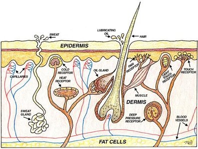
The cutaneous membrane, commonly known as skin, is the body's outer boundary and is classified as a dry membrane. It consists of keratinized stratified squamous epithelium and an underlying dermis composed mostly of dense connective tissue.
Keratinized stratified squamous epithelium: Provides a tough, water-resistant barrier.
Dermis: Dense connective tissue supports and nourishes the epidermis.
Function: Protects against mechanical, chemical, bacterial, UV, thermal damage, and desiccation.
Mucous Membranes
Structure and Function
Mucous membranes line all body cavities that open to the exterior. They are adapted for absorption and secretion and consist of different epithelial types depending on location.
Stratified squamous epithelium: Found in the mouth and esophagus.
Simple columnar epithelium: Found in the rest of the digestive tract.
Function: Protects, absorbs, and secretes substances.
Serous Membranes
Structure and Function
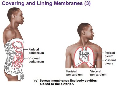
Serous membranes line body cavities closed to the exterior and consist of simple squamous epithelium over areolar connective tissue. They are separated by serous fluid, which reduces friction.
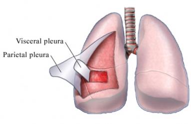
Visceral layer: Covers the outside of organs.
Parietal layer: Lines the wall of the ventral body cavity.
Serous fluid: Lubricates surfaces to prevent friction.
Serous Membrane Types
Peritoneum: Lines the abdominal cavity.
Pleura: Surrounds the lungs.
Pericardium: Surrounds the heart.
Connective Tissue Membranes
Synovial Membranes
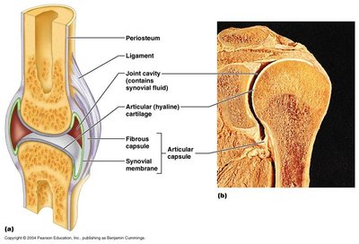
Synovial membranes are composed entirely of connective tissue and line the capsules surrounding joints. They secrete synovial fluid, which lubricates the joint.
Function: Reduces friction and nourishes articular cartilage.
Location: Found in movable joints such as the knee, elbow, and shoulder.
Integumentary System
Components
The integumentary system includes the skin and its derivatives: sweat glands, oil glands, hair, and nails.
Skin: Cutaneous membrane.
Sweat glands: Regulate temperature and excrete waste.
Oil glands: Lubricate skin and hair.
Hair: Provides protection and sensory input.
Nails: Protect fingertips and enhance sensation.
Functions of Skin
The skin performs several vital functions:
Protection: Shields deeper tissues from mechanical, chemical, bacterial, UV, thermal damage, and desiccation.
Temperature regulation: Aids in heat loss or retention as controlled by the nervous system.
Excretion: Removes urea and uric acid.
Vitamin D synthesis: Initiates production of vitamin D upon exposure to sunlight.
Skin Structure
Layers of the Skin
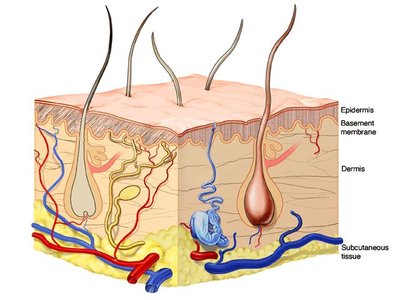
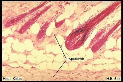
The skin is composed of three main layers:
Epidermis: Outermost layer, avascular, consists of keratinized stratified squamous epithelium.
Dermis: Dense connective tissue, contains blood vessels, nerves, and skin appendages.
Hypodermis: Deep to dermis, mostly adipose tissue, anchors skin to underlying organs.
Layers of the Epidermis
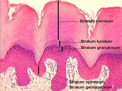
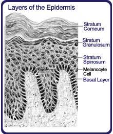
The epidermis consists of several distinct layers, each with specific functions.
Stratum basale (germinativum): Deepest layer, site of cell division.
Stratum spinosum: Provides strength and flexibility.
Stratum granulosum: Cells begin to die and accumulate granules.
Stratum lucidum: Present only in thick, hairless skin (palms, soles).
Stratum corneum: Outermost layer, dead cells filled with keratin.
Skin Color Determinants
Melanin
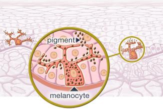
Melanin is a pigment produced by melanocytes, giving skin its color from yellow to brown to black. The amount produced depends on exposure to sunlight.
Function: Protects against UV radiation.
Location: Found in the stratum basale.
Other Color Determinants
Carotene: Orange-yellow pigment from vegetables.
Hemoglobin: Red coloring from blood cells in dermal capillaries; oxygen content affects redness.
Alterations in Skin Color
Redness (erythema): Caused by embarrassment, inflammation, hypertension, fever, or allergy.
Pallor (blanching): Due to emotional stress, anemia, low blood pressure, impaired blood flow.
Jaundice (yellowing): Indicates liver disorder.
Bruises: Result from blood escaping vessels and clotting under the skin.
Dermis
Structure and Function
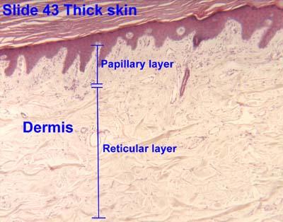
The dermis is composed of two layers:
Papillary layer: Upper dermal region, contains dermal papillae, pain and touch receptors.
Reticular layer: Deepest skin layer, contains blood vessels, sweat and oil glands, deep pressure receptors.
Dermal Structure
Collagen fibers: Provide toughness.
Elastic fibers: Allow stretch and recoil.
Blood vessels: Aid in temperature regulation.
Skin Appendages
Cutaneous Glands
Sebaceous (oil) glands: Produce sebum, lubricate skin, prevent brittle hair, kill bacteria. Ducts empty into hair follicles or onto skin surface. Activated at puberty.
Sweat (sudoriferous) glands: Produce sweat, widely distributed. Two types: eccrine (open to skin surface, clear sweat) and apocrine (empty into hair follicles, milky/yellowish sweat).
Sweat Composition and Function
Composition: Mostly water, salts, vitamin C, metabolic waste, fatty acids and proteins (apocrine only).
Function: Dissipates heat, excretes waste, inhibits bacterial growth due to acidity.
Hair
Produced by hair follicle: Keratinized cells, melanocytes provide pigment.
Growth: Occurs in the hair bulb.
Associated Hair Structures
Hair follicle: Surrounds and nourishes hair root.
Arrector pili muscle: Smooth muscle that pulls hair upright when cold or frightened.
Sebaceous gland: Lubricates hair.
Sudoriferous gland: Associated with hair follicle.
Nails
Structure: Scale-like modifications of the epidermis, heavily keratinized, colorless due to lack of pigment.
Growth: Occurs beneath the nail bed.
Parts: Free edge, body, root, cuticle, nail matrix, nail bed.
Table: Nail Structure
Part | Description |
|---|---|
Free edge | Distal portion, not attached |
Body | Visible attached portion |
Root | Embedded in skin |
Cuticle | Proximal nail fold projecting onto nail body |
Nail matrix | Responsible for growth |
Nail bed | Underlying area supporting nail |
Summary Table: Layers of the Skin
Layer | Location | Main Function |
|---|---|---|
Epidermis | Outer | Protection, water resistance |
Dermis | Middle | Support, nourishment, sensation |
Hypodermis | Deepest | Anchors skin, insulation, energy storage |
Key Terms
Keratin: Protein that hardens and waterproofs skin.
Melanocyte: Cell that produces melanin pigment.
Desmosome: Cell junction providing mechanical strength.
Dermal papillae: Projections increasing surface area for exchange.
Sudoriferous gland: Sweat gland.
Sebaceous gland: Oil gland.
Additional info:
Vitamin D synthesis in skin is initiated by UV light, which converts 7-dehydrocholesterol to cholecalciferol.
Keratinocytes are the most abundant cell type in the epidermis.
Collagen and elastin fibers in the dermis provide structural integrity and elasticity.
