 뒤로
Skin and Body Membranes: The Integumentary System
스터디 가이드 - 스마트 노트
 자료에 맞춘 맞춤형 노트, 핵심 정의, 예시, 맥락을 확장해 제공합니다.
Skin and Body Membranes: The Integumentary System
Classification of Body Membranes
Body membranes serve to cover surfaces, line body cavities, and form protective sheets around organs. They are classified based on tissue type into two major groups: epithelial membranes and connective tissue membranes.
Epithelial Membranes: Include cutaneous, mucous, and serous membranes.
Connective Tissue Membranes: Include synovial membranes.
Epithelial Membranes
Epithelial membranes are also known as covering and lining membranes. They are considered simple organs because they consist of both an epithelial layer and an underlying connective tissue layer.
Cutaneous Membrane (Skin): The skin is a dry membrane exposed to air, composed of a superficial epidermis (keratinized stratified squamous epithelium) and an underlying dermis (dense irregular connective tissue).
Mucous Membranes: Moist membranes lining body cavities that open to the exterior, such as the respiratory and digestive tracts. They consist of an epithelium (stratified squamous or simple columnar) and a loose connective tissue called lamina propria. Not all mucosae secrete mucus; some are adapted for absorption or secretion.
Serous Membranes: Line closed compartments in the ventral body cavity. Occur in pairs separated by serous fluid. The parietal layer lines the cavity wall, while the visceral layer covers the organ. Composed of simple squamous epithelium and areolar connective tissue. Examples include peritoneum (abdomen), pleurae (lungs), and pericardia (heart).
Connective Tissue Membranes
Synovial membranes are composed of loose areolar connective tissue and contain no epithelial cells. They line fibrous capsules surrounding joints, bursae, and tendon sheaths, providing a smooth surface and secreting lubricating fluid to cushion organs during movement.
The Integumentary System
The integumentary system, also known as the integument, consists of the skin and its appendages (sweat and oil glands, nails, hair). It performs various protective functions.
Insulates and cushions deeper organs
Protects against mechanical, chemical, thermal, UV, and bacterial damage
Prevents water loss
Regulates heat loss via capillary networks and sweat glands
Acts as a mini-excretory system (sweat removes urea, salts, water)
Manufactures proteins for immunity and vitamin D synthesis
Produces acidic secretions (acid mantle) to protect against pathogens
Synthesis of Vitamin D
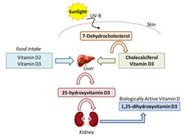
UVB radiation from sunlight converts 7-dehydrocholesterol in the skin to pre-cholecalciferol. Heat rearranges it to form vitamin D3 (cholecalciferol), which travels to the liver and is converted to 25-hydroxyvitamin D3 (calcidiol). In the kidneys, it is further converted to 1,25-dihydroxyvitamin D3 (calcitriol), the biologically active form.
Structure of the Skin
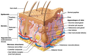
The skin is composed of two main tissue types: the epidermis (stratified squamous epithelium) and the dermis (dense irregular connective tissue). Beneath the dermis lies the hypodermis (subcutaneous tissue), primarily adipose tissue, which anchors the skin and provides insulation and shock absorption.
Epidermis: Avascular, composed mainly of keratinocytes connected by desmosomes. Keratinization makes the epidermis tough. Contains up to five layers (strata): stratum basale, stratum spinosum, stratum granulosum, stratum lucidum (in thick skin only), and stratum corneum.
Dermis: Strong, stretchy connective tissue with two regions: papillary (areolar connective tissue, dermal papillae, pain/touch receptors, fingerprints) and reticular (dense irregular connective tissue, blood vessels, glands, deep pressure receptors).
Hypodermis: Adipose tissue, not part of the skin but anchors it and stores nutrients.
Melanin and Skin Colour
Melanin is a pigment produced by melanocytes, primarily in the stratum basale. It ranges in color from yellow to brown to black and contributes to skin color.
Appendages of the Skin
Skin appendages include sweat glands, oil glands, hair, hair follicles, and nails. These structures are located in the dermis but originate from epidermal cells and play roles in homeostasis.
Homeostatic Imbalances of Skin
The skin can develop over 1,000 different ailments, including infections, allergies, burns, and cancer.
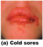
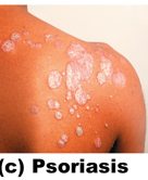
Infections and Allergies: Examples include athlete’s foot, contact dermatitis, cold sores, and psoriasis.
Burns: Tissue damage caused by heat, electricity, UV radiation, or chemicals. Burns are classified by degree:
Degree | Depth | Symptoms |
|---|---|---|
First | Superficial (epidermis) | Red, swollen |
Second | Epidermis + superficial dermis | Red, painful, blistered |
Third | Full-thickness (epidermis + dermis) | Painless, blanched or black, requires grafts |
Fourth | Extends to deeper tissues | Dry, leathery, may require amputation |
Critical Burn Criteria: Over 30% body with second-degree burns, over 10% with third/fourth-degree burns, burns of face/hands/feet/genitals, airway involvement, circumferential burns.
Skin Cancer
Skin cancer is the most common form of cancer in humans. The main risk factor is overexposure to UV radiation. Tumors can be benign (not spread) or malignant (invaded other areas).
Summary Table: Types of Body Membranes
Type | Location | Structure | Function |
|---|---|---|---|
Cutaneous | Skin | Keratinized stratified squamous epithelium + dense irregular connective tissue | Protection |
Mucous | Body cavities open to exterior | Epithelium + lamina propria | Absorption, secretion |
Serous | Closed body cavities | Simple squamous epithelium + areolar connective tissue | Lubrication |
Synovial | Joints, bursae, tendon sheaths | Areolar connective tissue | Lubrication, cushioning |
Key Equations
Vitamin D Synthesis:
Additional info:
Dermal papillae patterns are genetically determined and form unique fingerprints.
The acid mantle is a protective barrier against pathogens.
Burn severity is estimated using the rule of nines, dividing the body into areas representing 9% each.
