 뒤로
Smooth Muscle: Structure, Function, and Developmental Aspects
스터디 가이드 - 스마트 노트
 자료에 맞춘 맞춤형 노트, 핵심 정의, 예시, 맥락을 확장해 제공합니다.
Microscopic Structure of Smooth Muscle
General Characteristics
Smooth muscle is a type of muscle tissue found primarily in the walls of hollow organs, except the heart. It is composed of spindle-shaped fibers that are thin and short compared to skeletal muscle fibers, with a diameter of 5–10 µm and a length of 30–200 µm. Each fiber contains a single nucleus and lacks striations, distinguishing it from skeletal and cardiac muscle. Smooth muscle lacks connective tissue sheaths, containing only endomysium.
Spindle-shaped fibers: Thin, short, and single-nucleated.
No striations: Smooth appearance under the microscope.
Endomysium only: No perimysium or epimysium.
Organization in Hollow Organs
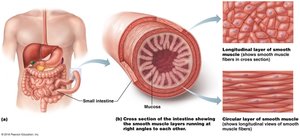
Smooth muscle is typically organized into two sheets within the walls of hollow organs:
Longitudinal layer: Fibers run parallel to the organ's long axis; contraction shortens the organ.
Circular layer: Fibers encircle the organ; contraction constricts the lumen.
Peristalsis: Alternating contractions and relaxations mix and propel substances through the lumen.
Innervation and Junctions
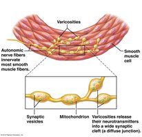
Smooth muscle lacks neuromuscular junctions. Instead, it is innervated by autonomic nerve fibers that contain varicosities—bulbous swellings storing and releasing neurotransmitters into a wide synaptic cleft called a diffuse junction.
Varicosities: Store and release neurotransmitters.
Diffuse junction: Wide synaptic cleft for neurotransmitter release.
Cellular Structure and Calcium Handling
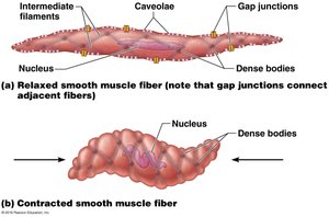
Smooth muscle does not contain sarcomeres, myofibrils, or T tubules. The sarcoplasmic reticulum (SR) is less developed, and most calcium for contraction enters from the extracellular space via caveolae—pouch-like infoldings in the sarcolemma containing Ca2+ channels.
Caveolae: Infoldings with Ca2+ channels for rapid influx.
Calcium removal: Contraction ends when Ca2+ is actively transported into the SR and out of the cell.
Filament Arrangement and Dense Bodies
Thick filaments are fewer in smooth muscle (ratio 1:13) compared to skeletal muscle (1:2). Myosin heads are found along the entire length, and there is no troponin complex. Instead, calmodulin binds Ca2+. Filaments are arranged diagonally, causing corkscrew contractions. Intermediate filaments and dense bodies form a lattice-like network, anchoring thin filaments and facilitating synchronous contractions.
Calmodulin: Binds Ca2+ instead of troponin.
Dense bodies: Anchor filaments and correspond to Z discs in skeletal muscle.
Contraction of Smooth Muscle
Mechanism and Regulation
Smooth muscle exhibits slow, synchronized contractions due to gap junctions. Some fibers are self-excitatory and act as pacemakers. Contraction is similar to skeletal muscle in that actin and myosin interact via the sliding filament mechanism, and ATP is required. However, most Ca2+ comes from the extracellular space, and Ca2+ binds to calmodulin, not troponin.
Gap junctions: Allow action potentials to spread between fibers.
Pacemaker cells: Set contraction pace for muscle sheets.
Sliding filament mechanism: Actin and myosin interaction.
Excitation-Contraction Coupling Sequence
The sequence of events in smooth muscle contraction involves several steps:
Calcium ions (Ca2+) enter the cytosol from the extracellular fluid or scant SR.
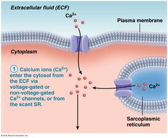
Ca2+ binds to and activates calmodulin.
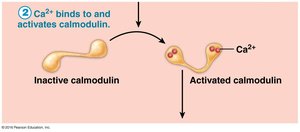
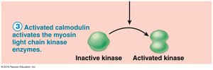
Activated calmodulin activates myosin light chain kinase enzymes.
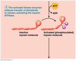
Activated kinase enzymes catalyze phosphorylation of myosin, activating myosin ATPases.
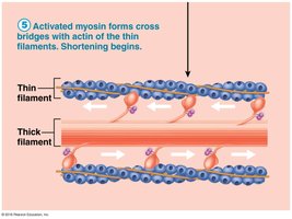
Activated myosin forms cross bridges with actin, initiating contraction.
Relaxation and Energy Efficiency
Relaxation requires Ca2+ detachment from calmodulin, active transport of Ca2+ into the SR and extracellularly, and dephosphorylation of myosin. Smooth muscle contracts and relaxes 30 times slower than skeletal muscle but maintains contractions for prolonged periods with minimal energy cost due to slower ATPases and latch mechanisms.
Smooth muscle tone: Maintains moderate contraction without fatigue.
Energy requirements: Low; ATP produced mainly via aerobic respiration.
Regulation by Nerves, Hormones, and Local Factors
Contraction is controlled by neural, hormonal, or local chemical stimuli. Neurotransmitter binding can stimulate or inhibit contraction depending on receptor type. Hormones and local chemicals (e.g., CO2, pH, O2) can also modulate contraction.
Neural regulation: Action potential and Ca2+ increase.
Hormonal regulation: Example: gastrin stimulates stomach contraction.
Local chemical regulation: Factors like pH and O2 affect contraction.
Special Features
Smooth muscle responds to stretch with a stress-relaxation response, adapting to new lengths while retaining contractility. It can stretch more and generate more tension than skeletal muscle, contracting over a length change of ~150%.
Types of Smooth Muscle
Unitary (Visceral) Smooth Muscle
Found in all hollow organs except the heart, unitary smooth muscle is arranged in opposing sheets, innervated by varicosities, and contracts as a unit due to gap junctions. It responds to various chemical stimuli and often exhibits rhythmic, spontaneous action potentials.
Multiunit Smooth Muscle
Located in large airways, arteries, arrector pili muscles, and the iris, multiunit smooth muscle consists of independent fibers with few gap junctions. It is richly innervated, contracts in a graded manner, and is controlled by the autonomic nervous system and hormones.
Comparison of Muscle Types
Structural and Functional Differences
The following tables summarize the key differences between skeletal, cardiac, and smooth muscle:
Characteristic | Skeletal | Cardiac | Smooth |
|---|---|---|---|
Body location | Attached to bones or facial muscles | Walls of the heart | Walls of hollow organs (except heart) |
Cell shape and appearance | Single, very long, cylindrical, multinucleate, striated | Branching chains, striated, 1-2 nuclei | Single, fusiform, uninucleate, no striations |
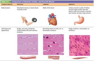
Characteristic | Skeletal | Cardiac | Smooth |
|---|---|---|---|
Connective tissue components | Epimysium, perimysium, endomysium | Endomysium attached to fibrous skeleton | Endomysium only |
Presence of myofibrils | Yes | Yes, irregular | No |
Presence of T tubules | Yes | Yes, fewer | No |
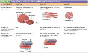
Characteristic | Skeletal | Cardiac | Smooth |
|---|---|---|---|
Elaborate sarcoplasmic reticulum | Yes | Less than skeletal | Equivalent to cardiac |
Presence of gap junctions | No | Yes, at intercalated discs | Yes, in unitary muscle |
Cellular regulation | Voluntary | Involuntary | Involuntary |
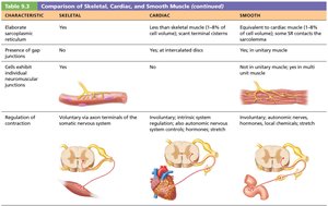
Characteristic | Skeletal | Cardiac | Smooth |
|---|---|---|---|
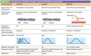
Source of Ca2+ | Sarcoplasmic reticulum | SR and extracellular fluid | SR and extracellular fluid |
Regulatory proteins | Troponin on actin | Troponin on actin | Calmodulin in cytosol |
Speed of contraction | Slow to fast | Slow | Very slow |
Developmental Aspects of Muscle Tissue
Embryonic Development
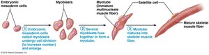
All muscle tissues develop from embryonic mesoderm cells called myoblasts. Skeletal muscle cells become multinucleated by fusion of myoblasts, while cardiac and smooth muscle myoblasts do not fuse but develop gap junctions. Cardiac muscle begins functioning early in embryonic development.
Regeneration and Growth
Skeletal muscle: Satellite cells have limited regenerative ability.
Cardiac muscle: Limited regeneration; mostly replaced by connective tissue.
Smooth muscle: Regenerates throughout life.
Hypertrophy: Muscle fibers can lengthen and thicken in children and adults.
Neuromuscular Coordination and Muscle Mass
Infant movements: Reflexive and uncoordinated; neuromuscular control improves with age.
Sex differences: Males have greater muscle mass due to testosterone, but strength per unit mass is equal in both sexes.
Aging and Muscle Disorders
Sarcopenia: Age-related loss of muscle mass begins at age 30; regular exercise can reverse effects.
Atherosclerosis: Aging cardiovascular system may lead to blocked arteries and muscle pain.
Homeostatic Imbalance: Muscular Dystrophy
Duchenne Muscular Dystrophy (DMD)
Muscular dystrophy is a group of inherited muscle-destroying diseases, with DMD being the most common and severe. It is caused by a defective gene for dystrophin, a protein that stabilizes the sarcolemma. The disease is sex-linked, carried by females and expressed in males.
Dystrophin: Links cytoskeleton to extracellular matrix; stabilizes sarcolemma.
Pathology: Sarcolemma tears, excess Ca2+ entry, fiber damage, inflammation, and loss of regenerative ability.
Symptoms: Frequent falls, muscle weakness, respiratory failure.
Treatment: No cure; prednisone and immunosuppressants may delay progression.
Additional info: DMD is diagnosed in boys between ages 2 and 7, and muscle mass declines progressively.
