 뒤로
muscle 2
스터디 가이드 - 스마트 노트
 자료에 맞춘 맞춤형 노트, 핵심 정의, 예시, 맥락을 확장해 제공합니다.
Muscle Structure and Somatic Motor Control
Overview of Skeletal Muscle Anatomy
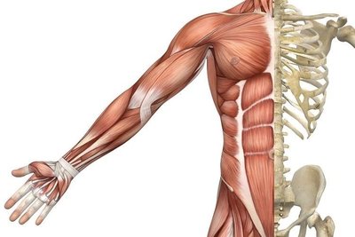
Skeletal muscle is composed of bundles of muscle fibers, each innervated by motor neurons. The somatic motor division of the nervous system controls voluntary muscle contraction, allowing precise and powerful movements.
Muscle fibers are elongated cells containing contractile proteins.
Motor neurons transmit signals from the central nervous system to muscle fibers.
Motor unit: A single motor neuron and all the muscle fibers it innervates.
The number of muscle fibers per motor unit varies: fewer for precise movements, more for powerful contractions.
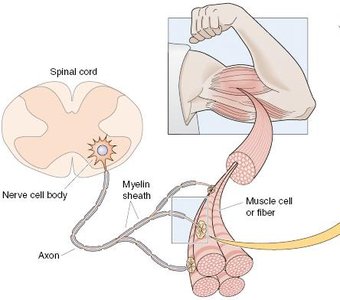
Somatic Motor Neuron Pathway
Somatic motor neurons originate in the spinal cord and extend their axons to skeletal muscle fibers. The pathway includes upper motor neurons (brain to spinal cord) and alpha (lower) motor neurons (spinal cord to muscle).
Upper motor neuron: Brain to spinal cord or brainstem.
Alpha motor neuron: Spinal cord to muscle.
Axons are large and myelinated, allowing rapid signal transmission (15–120 m/sec).
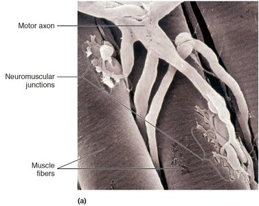
Neuromuscular Junction
Structure and Function
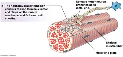
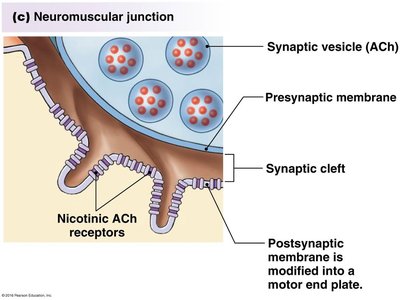
The neuromuscular junction (NMJ) is the synapse between a motor neuron and a skeletal muscle fiber. It is essential for converting neural signals into muscle contraction.
Three main components: presynaptic motor neuron (with synaptic vesicles), synaptic cleft, and postsynaptic muscle membrane (motor end plate).
Motor neuron vesicles contain acetylcholine (ACh).
Muscle sarcolemma contains nicotinic acetylcholine receptors.
Junctional folds on the sarcolemma increase surface area for receptor density.
Synaptic Transmission at the NMJ
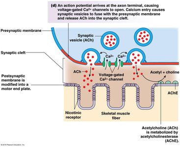
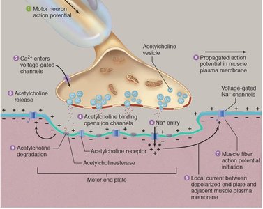
When an action potential reaches the axon terminal, voltage-gated Ca2+ channels open, allowing Ca2+ influx. This triggers the release of ACh into the synaptic cleft, which binds to nicotinic receptors on the motor end plate, generating an excitatory end-plate potential (EPP).
Nicotinic ACh receptor: Ligand-gated ion channel permeable to Na+ and K+.
Opening requires two ACh molecules.
Na+ entry depolarizes the sarcolemma, initiating an action potential.
Acetylcholine Recycling and Termination
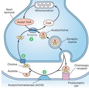
After neural transmission, acetylcholine must be removed from the synaptic cleft to prevent continuous stimulation. Acetylcholinesterase breaks down ACh into acetate and choline. Choline is transported back into the neuron and combined with acetyl CoA to synthesize new ACh.
Acetylcholinesterase: Enzyme that hydrolyzes ACh.
Choline acetyltransferase: Enzyme that synthesizes ACh from choline and acetyl CoA.
Disorders of Neuromuscular Transmission
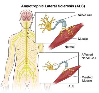
Amyotrophic Lateral Sclerosis (ALS)
ALS is a neurodegenerative disease affecting motor neurons, leading to muscle atrophy and weakness. Both upper and lower motor neurons can degenerate, resulting in eventual respiratory failure.
10% of cases are genetically inherited (familial).
25% of familial cases involve mutations in superoxide dismutase genes (antioxidant defense).
Symptoms: Progressive muscle weakness, atrophy, and loss of voluntary movement.
Myasthenia Gravis
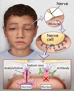
Myasthenia gravis is an autoimmune disorder where antibodies target ACh receptors, impairing neuromuscular transmission and causing muscle weakness.
Can affect specific muscles (e.g., extraocular) or be generalized.
Antibodies block or destroy ACh receptors, reducing muscle activation.
Treatment: Acetylcholinesterase inhibitors or immunosuppressants.
Excitation-Contraction Coupling
Propagation of Action Potentials
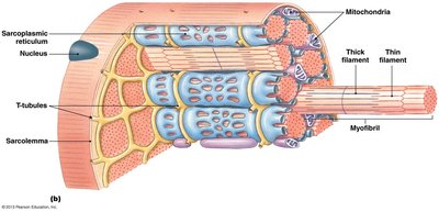
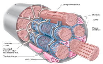
Action potentials generated at the motor end plate propagate along the sarcolemma and into the muscle fiber via transverse (T-) tubules. This ensures rapid and uniform activation of the muscle.
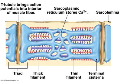
T-tubules: Invaginations of the sarcolemma that conduct action potentials deep into the muscle fiber.
Sarcoplasmic reticulum (SR): Specialized organelle for Ca2+ storage and release.
Triad Structure and Calcium Release
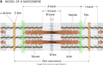
T-tubules are closely associated with terminal cisternae of the SR, forming a triad at the A and I band junctions of each sarcomere. This arrangement facilitates efficient Ca2+ release during muscle contraction.
Triad: Two terminal cisternae and one T-tubule.
SR releases Ca2+ in response to action potentials.
Excitation-Contraction Coupling Mechanism
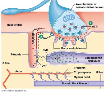
The process by which electrical excitation triggers muscle contraction is known as excitation-contraction coupling. The ultimate signal is a rise in intracellular Ca2+, which binds to troponin and initiates cross-bridge cycling.
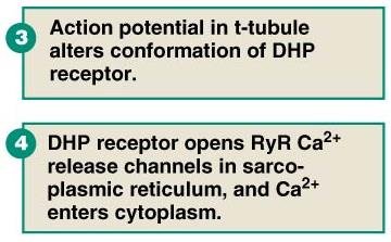
Action potential in T-tubule alters conformation of DHP receptor (L-type Ca2+ channel).
DHP receptor opens RyR Ca2+ release channels in SR, allowing Ca2+ to enter cytoplasm.
Ca2+ binds to troponin, shifting tropomyosin and exposing actin binding sites for myosin.
Component | Function |
|---|---|
DHP receptor | Voltage-sensitive L-type Ca2+ channel in T-tubule |
RyR receptor | Ca2+ release channel in SR |
Troponin | Ca2+ binding protein, initiates contraction |
Tropomyosin | Regulates access to actin binding sites |
