 뒤로
Spinal Cord, Spinal Meninges, Spinal Nerves, and Nerve Plexuses: Study Guide
스터디 가이드 - 스마트 노트
 자료에 맞춘 맞춤형 노트, 핵심 정의, 예시, 맥락을 확장해 제공합니다.
Spinal Cord: Gross and Sectional Anatomy
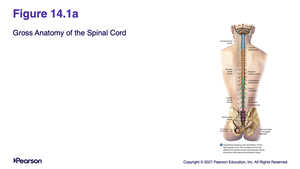
Gross Anatomy of the Spinal Cord
The spinal cord is a major component of the central nervous system, extending from the brainstem down through the vertebral column. It is responsible for transmitting neural signals between the brain and the rest of the body, and for coordinating reflexes.
Location: Runs within the vertebral canal, protected by vertebrae.
Regions: Cervical, thoracic, lumbar, sacral, and coccygeal.
Functions: Conduction of sensory and motor information; integration of reflexes.
Sectional Organization of the Spinal Cord
The spinal cord is organized into gray and white matter, each with distinct functions. Gray matter contains neuron cell bodies, while white matter consists of myelinated axons.
Gray Matter: Organized into horns (anterior, lateral, posterior) responsible for motor and sensory processing.
White Matter: Organized into columns (anterior, lateral, posterior) for signal transmission.
Functional Areas: Somatic and visceral sensory, motor regions.
Spinal Meninges
Overview of Spinal Meninges
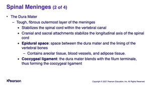
Spinal meninges are specialized membranes that surround the spinal cord, providing protection, stability, and shock absorption. They are continuous with the cranial meninges and consist of three layers.
Dura mater: Tough, fibrous outermost layer.
Arachnoid mater: Middle layer.
Pia mater: Innermost layer, adjacent to spinal tissue.
Dura Mater
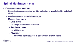
The dura mater is the tough, fibrous outermost layer of the meninges. It stabilizes the spinal cord within the vertebral canal and forms attachments at the cranial and sacral ends.
Epidural space: Space between dura mater and vertebral bones; contains areolar tissue, blood vessels, and adipose tissue.
Coccygeal ligament: Dura mater blends with filum terminale to form this ligament.
Arachnoid Mater
The arachnoid mater is the middle meningeal layer, separated from the pia mater by the subarachnoid space. Cerebrospinal fluid flows within this space, providing cushioning and nutrient transport.
Subarachnoid space: Contains cerebrospinal fluid.
Arachnoid trabeculae: Bundles of fibers connecting arachnoid to pia mater.
Subdural space: Found in cadavers, not in living people.
Pia Mater
The pia mater is the deepest meningeal layer, firmly attached to the brain and spinal cord tissue. It contains blood vessels and forms extensions called denticulate ligaments, which stabilize the spinal cord.
Denticulate ligaments: Prevent side-to-side and downward movement.
Filum terminale: Formed at the inferior tip of the conus medullaris.
Spinal Nerves
Overview and Numbering of Spinal Nerves
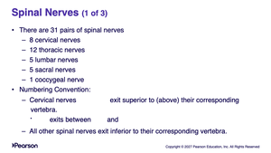
Spinal nerves are mixed nerves that emerge from the spinal cord, supplying motor and sensory information to the body. There are 31 pairs, each associated with a specific region.
8 cervical nerves
12 thoracic nerves
5 lumbar nerves
5 sacral nerves
1 coccygeal nerve
Numbering convention: Cervical nerves exit superior to their corresponding vertebrae; all other spinal nerves exit inferior.
Structure of Peripheral Nerves
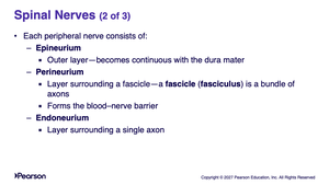
Each peripheral nerve consists of three connective tissue layers:
Epineurium: Outer layer, continuous with dura mater.
Perineurium: Surrounds fascicles (bundles of axons); forms blood-nerve barrier.
Endoneurium: Surrounds individual axons.
Peripheral Distribution and Branches
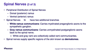
Spinal nerves branch into dorsal (posterior) and ventral (anterior) rami, supplying different regions. They also have additional branches called rami communicantes.
White ramus communicans: Carries myelinated preganglionic axons to the sympathetic ganglion.
Gray ramus communicans: Carries unmyelinated postganglionic axons back to the spinal nerve.
Dermatomes: Specific regions of skin supplied by spinal nerves.
Nerve Plexuses
Overview of Nerve Plexuses
Nerve plexuses are interwoven networks of nerves formed during embryonic development from the ventral rami of adjacent spinal nerves. They innervate fused muscle groups and are essential for coordinated movement.
Cervical plexus
Brachial plexus
Lumbar plexus
Sacral plexus
Sometimes lumbar and sacral are combined as the lumbosacral plexus.
Cervical Plexus
The cervical plexus is composed of ventral rami of spinal nerves and some fibers from other nerves. It is formed from cutaneous and muscular branches that innervate the head, neck, and chest.
Cutaneous branches: Supply skin of head, neck, chest.
Muscular branches: Supply muscles of neck and diaphragm.
Brachial Plexus
The brachial plexus innervates the pectoral girdle and upper limbs. It originates from the ventral rami of spinal nerves and is organized into roots, trunks, divisions, cords, and nerves.
Trunks: Superior, middle, and inferior
Divisions: Anterior and posterior
Cords: Posterior, lateral, and medial
Nerves: Major peripheral nerves of the upper limb
Summary Table: Spinal Meninges
The following table summarizes the three layers of spinal meninges and their key features:
Layer | Location | Key Features |
|---|---|---|
Dura mater | Outermost | Tough, fibrous; stabilizes spinal cord; forms coccygeal ligament |
Arachnoid mater | Middle | Contains subarachnoid space; cerebrospinal fluid; arachnoid trabeculae |
Pia mater | Innermost | Contains blood vessels; forms denticulate ligaments and filum terminale |
Summary Table: Nerve Plexuses
This table summarizes the four main nerve plexuses and their regions of innervation:
Plexus | Region Innervated |
|---|---|
Cervical | Head, neck, chest |
Brachial | Pectoral girdle, upper limbs |
Lumbar | Lower back, anterior and medial thigh |
Sacral | Pelvis, posterior thigh, leg, foot |
