 뒤로
Structural Organization and Function of Skeletal Muscle Fibers
스터디 가이드 - 스마트 노트
 자료에 맞춘 맞춤형 노트, 핵심 정의, 예시, 맥락을 확장해 제공합니다.
Skeletal Muscle Fiber Structure
Distinctive Features of Skeletal Muscle Fibers
Skeletal muscle fibers are specialized cells designed for contraction and movement. Unlike typical cells, they are large, multinucleate, and contain unique structures that facilitate their function.
Size: Muscle fibers can reach up to 60 cm in length and 100 μm in diameter.
Multinucleate: Each fiber contains hundreds of nuclei beneath the sarcolemma.
Components: Key structures include the sarcolemma, T tubules, myofibrils, and sarcoplasmic reticulum.
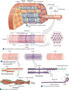
The Sarcolemma and Transverse Tubules (T Tubules)
The sarcolemma is the plasma membrane of the muscle fiber, surrounding the sarcoplasm. Openings in the sarcolemma lead to T tubules, which are filled with extracellular fluid and form passageways through the fiber.
T tubules: Allow electrical impulses to reach deep into the muscle fiber, ensuring simultaneous contraction throughout the cell.
Function: Electrical impulses travel along T tubules, triggering contraction by altering the chemical environment inside the fiber.
Myofibrils and Myofilaments
Myofibrils are cylindrical structures within muscle fibers, composed of bundles of myofilaments (actin and myosin). Each fiber contains hundreds to thousands of myofibrils.
Actin: Found in thin filaments.
Myosin: Found in thick filaments.
Contraction: Myofibrils shorten, causing the entire muscle fiber to contract.
Energy: Mitochondria and glycogen granules provide ATP for contraction.
Sarcoplasmic Reticulum (SR) and Terminal Cisternae
The sarcoplasmic reticulum is a specialized smooth endoplasmic reticulum that surrounds each myofibril. It stores calcium ions, which are essential for muscle contraction.
Terminal cisternae: Expanded chambers of the SR, located on either side of T tubules, forming a triad.
Calcium storage: Calcium ions are actively transported into terminal cisternae and released into the sarcoplasm to initiate contraction.
Sarcomere Structure and Function
Organization of the Sarcomere
The sarcomere is the smallest functional unit of a muscle fiber, responsible for contraction. It consists of repeating arrangements of thick and thin filaments.
Z lines: Boundaries of each sarcomere; thin filaments are attached here.
A band: Dark region containing thick filaments.
I band: Light region containing thin filaments and the Z line.
Zone of overlap: Area where thick and thin filaments overlap.
M line: Proteins connecting central portions of thick filaments.
H band: Region containing only thick filaments, including the M line.
Titin: Protein strands that maintain alignment of filaments.
Thin and Thick Filaments
Thin filaments are composed of actin, tropomyosin, and troponin. Thick filaments are made of myosin molecules, each with a tail and globular head.
Actin: Contains active sites for myosin binding.
Tropomyosin: Covers active sites on actin in resting muscle.
Troponin: Holds tropomyosin in place; binds calcium to initiate contraction.
Myosin: Heads project outward and bind to actin during contraction.
Role of Calcium in Muscle Contraction
Calcium ions are crucial for muscle contraction. When released from the terminal cisternae, they bind to troponin, causing a conformational change that moves tropomyosin and exposes actin's active sites.
Calcium binding: Initiates the interaction between actin and myosin.
Source: Terminal cisternae of the sarcoplasmic reticulum.
Sliding Filament Theory and Cross-Bridge Cycling
Sliding Filament Theory
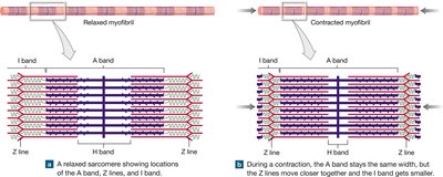
The sliding filament theory explains how muscle contraction occurs at the molecular level. Thin filaments slide toward the center of the sarcomere, alongside stationary thick filaments, resulting in shortening of the sarcomere.
During contraction: I bands and H bands decrease in size, Z lines move closer, A band remains unchanged.
Cross-bridges: Myosin heads bind to actin, pivot, detach, and repeat the cycle, pulling thin filaments inward.
Cross-Bridge Formation and Cycling
Cross-bridges are formed when myosin heads attach to actin's active sites. The cycle involves attachment, pivoting, detachment, and return, similar to pulling a rope.
Attach: Myosin head binds to actin.
Pivot: Head pivots, pulling actin toward the center.
Detach: Head releases actin.
Return: Head returns to original position, ready to repeat.
Calcium Metabolism and Muscle Function
Calcium's Role in the Body
Calcium is the most abundant mineral in the human body, essential for muscle contraction, bone health, and metabolic processes.
Vitamin D: Synthesized in the skin, modified by the liver, and converted by the kidneys to calcitriol, which is necessary for calcium absorption.
Bones: Serve as a mineral reserve for maintaining normal calcium and phosphate concentrations in body fluids.
Formula for Calcium Ion Transport:
Additional info: Calcium metabolism is tightly regulated by hormonal control, involving parathyroid hormone, calcitonin, and calcitriol.
