 뒤로
Study Guide: Brain and Cranial Nerves (Anatomy & Physiology)
스터디 가이드 - 스마트 노트
 자료에 맞춘 맞춤형 노트, 핵심 정의, 예시, 맥락을 확장해 제공합니다.
Major Regions of the Brain
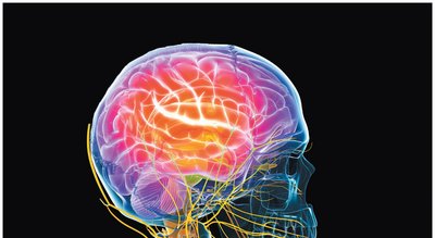
Cerebrum
The cerebrum is the largest part of the brain, responsible for conscious thought, intellectual functions, memory storage, and processing. It also regulates skeletal muscle contractions both consciously and subconsciously.
Gyri: Elevated ridges that increase surface area for neurons.
Sulci: Shallow depressions separating gyri.
Fissures: Deep grooves separating hemispheres and lobes.
Corpus callosum: Connects the two cerebral hemispheres.
Diencephalon
The diencephalon is a deep region attached to the cerebrum, consisting of the thalamus, hypothalamus, and epithalamus. It is involved in sensory relay, autonomic functions, and hormone production.
Thalamus: Relay and processing center for sensory information.
Hypothalamus: Controls emotions, autonomic functions, and hormone production; links nervous and endocrine systems.
Epithalamus: Contains the pineal gland (endocrine function).
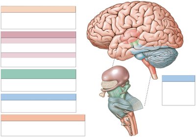
Brainstem
The brainstem includes the midbrain (mesencephalon), pons, and medulla oblongata. It is essential for relaying information and regulating vital functions.
Midbrain: Processes visual and auditory data, generates reflexive motor responses, maintains consciousness.
Pons: Relays sensory information, contains somatic and visceral motor centers.
Medulla oblongata: Relays sensory information, regulates cardiovascular, respiratory, and digestive activities.
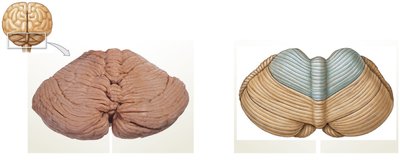
Cerebellum
The cerebellum coordinates complex somatic motor patterns and adjusts output of other motor centers. It is crucial for balance and equilibrium.
Arbor vitae: Branching white matter structure.
Purkinje cells: Large neurons in the cerebellar cortex.
Diencephalon: Thalamus and Hypothalamus
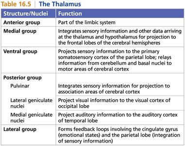
Thalamus
The thalamus is a major relay station for sensory and motor information. It consists of several nuclei, each with specific functions.
Anterior group: Part of the limbic system.
Medial group: Integrates sensory information for projection to the frontal lobes.
Ventral group: Projects sensory information to the somatosensory cortex; relays motor information.
Posterior group: Pulvinar integrates sensory info; lateral geniculate nuclei project visual info; medial geniculate nuclei project auditory info.
Lateral group: Forms feedback loops for emotional states and sensory integration.
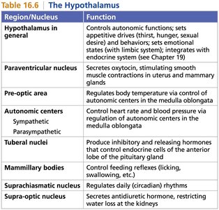
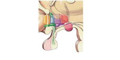
Hypothalamus
The hypothalamus is a control center for autonomic functions, emotional states, and hormone production. It connects to the pituitary gland and integrates nervous and endocrine systems.
Paraventricular nucleus: Secretes oxytocin for smooth muscle contraction.
Pre-optic area: Regulates body temperature.
Autonomic centers: Control heart rate and blood pressure.
Tuberal nuclei: Produce hormones for pituitary regulation.
Mammillary bodies: Control feeding reflexes.
Suprachiasmatic nucleus: Regulates circadian rhythms.
Supra-optic nucleus: Secretes antidiuretic hormone.
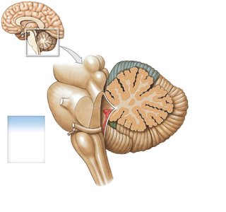
Cerebellum: Structure and Function
Anatomy of the Cerebellum
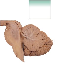
The cerebellum consists of two hemispheres, anterior and posterior lobes, and the vermis. It is located posterior to the pons and inferior to the cerebral hemispheres.
Folia: Folds of the cerebellar cortex.
Arbor vitae: White matter structure connecting cortex and nuclei.
Cerebellar peduncles: Superior, middle, and inferior peduncles link the cerebellum to other brain regions.
Functional Regions
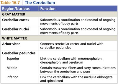
Cerebellar cortex: Subconscious coordination and control of movements.
Cerebellar nuclei: Subconscious coordination and control of movements.
Arbor vitae: Connects cortex and nuclei with peduncles.
Cerebellar peduncles: Superior links with mesencephalon, diencephalon, cerebrum; middle carries communication with pons; inferior links with medulla and spinal cord.
Cerebral Hemispheres and Lobes
Anatomy of the Cerebral Hemispheres
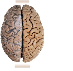
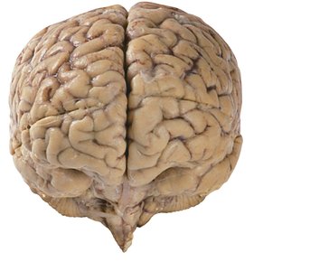
The cerebral hemispheres are separated by the longitudinal fissure and connected by the corpus callosum. The cortex is composed of gray matter, with gyri, sulci, and fissures increasing surface area.
Frontal lobe: Motor functions, reasoning, planning.
Parietal lobe: Sensory processing.
Temporal lobe: Auditory processing, memory.
Occipital lobe: Visual processing.
Functional Organization of the Cortex
The cortex is organized into primary areas (sensory and motor) and association areas (integration and interpretation). The central sulcus separates motor and sensory regions.
Primary motor cortex: Controls voluntary movements.
Primary somatosensory cortex: Receives sensory information.
Association areas: Integrate and interpret sensory/motor information.
Integrative Centers and Hemispheric Lateralization
Integrative centers process complex sensory stimuli and motor responses, including language, reasoning, and emotional context. Hemispheric lateralization refers to functional differences between the left and right hemispheres.
Wernicke's area: Language comprehension.
Broca's area: Speech production.
Prefrontal cortex: Learning, reasoning, emotional context.
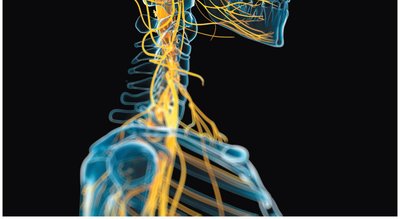
Cranial Nerves
Overview of Cranial Nerves
There are twelve pairs of cranial nerves, each with specific sensory, motor, or mixed functions. They are numbered according to their position along the brain.
Olfactory (I): Sensory, smell.
Optic (II): Sensory, vision.
Oculomotor (III): Motor, eye movement.
Trochlear (IV): Motor, eye movement.
Trigeminal (V): Both, facial sensation and mastication.
Abducens (VI): Motor, eye movement.
Facial (VII): Both, taste and facial expression.
Vestibulocochlear (VIII): Sensory, balance and hearing.
Glossopharyngeal (IX): Both, taste and swallowing.
Vagus (X): Both, visceral organ control.
Accessory (XI): Motor, neck muscles.
Hypoglossal (XII): Motor, tongue movement.
Mnemonic Devices
Names: On Occasion Our Trusty Truck Acts Funny Very Good Vehicle Any How
Functions: Some Say Marry Money But My Brother Says Big Brains Matter More
Clinical Note: Traumatic Brain Injury (TBI)
Types and Effects
Traumatic brain injuries result from harsh contact or jolts to the head. Types include concussions, chronic traumatic encephalopathy (CTE), subdural hematoma (venous bleed), and epidural hematoma (arterial bleed).
Concussion: Temporary loss of brain function.
CTE: Degenerative brain disease from repeated trauma.
Subdural hematoma: Blood between dura and arachnoid mater.
Epidural hematoma: Blood between skull and dura mater.
Summary Table: Thalamic Nuclei and Functions
Structure/Nuclei | Function |
|---|---|
Anterior group | Part of the limbic system |
Medial group | Integrates sensory information and other data arriving at the thalamus and hypothalamus for projection to the frontal lobes of the cerebral hemispheres |
Ventral group | Projects sensory information to the primary somatosensory cortex of the parietal lobe; relays information from cerebellum and basal nuclei to motor areas of cerebral cortex |
Posterior group | Pulvinar: Integrates sensory information for projection to association areas of cerebral cortex Lateral geniculate nuclei: Project visual information to the visual cortex of occipital lobe Medial geniculate nuclei: Project auditory information to the auditory cortex of temporal lobe |
Lateral group | Forms feedback loops involving the cingulate gyrus (emotional states) and the parietal lobe (integration of sensory information) |
Summary Table: Hypothalamic Nuclei and Functions
Region/Nucleus | Function |
|---|---|
Hypothalamus in general | Controls autonomic functions; sets appetitive drives (thirst, hunger, sexual desire) and behaviors; sets emotional states (with limbic system); integrates with endocrine system |
Paraventricular nucleus | Secretes oxytocin, stimulating smooth muscle contractions in uterus and mammary glands |
Pre-optic area | Regulates body temperature via control of autonomic centers in the medulla oblongata |
Autonomic centers | Control heart rate and blood pressure via regulation of autonomic centers in the medulla oblongata |
Tuberal nuclei | Produce inhibitory and releasing hormones that control endocrine cells of the anterior lobe of the pituitary gland |
Mammillary bodies | Control feeding reflexes (licking, swallowing, etc.) |
Suprachiasmatic nucleus | Regulates daily (circadian) rhythms |
Supra-optic nucleus | Secretes antidiuretic hormone, restricting water loss at the kidneys |
Summary Table: Cerebellar Regions and Functions
Region/Nucleus | Function |
|---|---|
Cerebellar cortex | Subconscious coordination and control of ongoing movements of body parts |
Cerebellar nuclei | Subconscious coordination and control of ongoing movements of body parts |
Arbor vitae | Connects cerebellar cortex and nuclei with cerebellar peduncles |
Superior cerebellar peduncle | Link the cerebellum with mesencephalon, diencephalon, and cerebrum |
Middle cerebellar peduncle | Contain transverse fibers and carry communications between the cerebellum and pons |
Inferior cerebellar peduncle | Link the cerebellum with the medulla oblongata and spinal cord |
Key Equations and Concepts
Neural Pathways: Sensory information is relayed from peripheral receptors to the thalamus, then projected to the appropriate cortical area for processing.
Motor Pathways: Motor commands originate in the cortex and are relayed through the brainstem and spinal cord to effectors.
Example: Visual information travels from the retina via the optic nerve (II), crosses at the optic chiasm, is relayed through the lateral geniculate nucleus of the thalamus, and reaches the occipital cortex for interpretation.
Additional info: The cerebellum's role in motor learning (muscle memory) and postural control is essential for coordinated movement and balance. The hypothalamus integrates nervous and endocrine functions, making it a key regulator of homeostasis.
