 뒤로
Study Guide: General and Special Senses in Human Anatomy
스터디 가이드 - 스마트 노트
 자료에 맞춘 맞춤형 노트, 핵심 정의, 예시, 맥락을 확장해 제공합니다.
Ch. 18: General and Special Senses
Introduction to Sensory Systems
The human body relies on sensory receptors to monitor internal and external conditions. Sensory information is processed as sensation (neural signals) and perception (conscious awareness). Senses are classified as general senses (widely distributed) and special senses (localized in specific organs).
General senses: Include temperature, pain, touch, pressure, vibration, and proprioception.
Special senses: Include smell (olfaction), taste (gustation), balance (equilibrium), hearing, and vision.
Receptors for special senses are found in specialized organs such as the eye and ear.
Receptor Properties and Classification
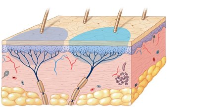
Sensory receptors exhibit specificity and are organized into receptive fields. The size of a receptive field affects the ability to localize a sensation.
Phasic receptors: Normally inactive, respond to changes in stimulus, provide information about intensity and rate of change.
Tonic receptors: Always active when stimulus is present, continuously send information to the CNS.
Adaptation: Reduction in sensitivity due to constant stimulation. Can be peripheral (receptor level) or central (CNS level).
Classification of General Sensory Receptors
General sensory receptors are scattered throughout the body and classified by location or stimulus type:
Exteroceptors: Monitor external environment.
Proprioceptors: Monitor body position.
Interoceptors: Monitor internal environment.
By stimulus type:
Nocioceptors: Pain receptors, free nerve endings, large receptive fields. Types: sensitive to extreme temperature, mechanical damage, or chemicals.
Thermoreceptors: Detect temperature changes within safe range.
Mechanoreceptors: Respond to physical distortion, contact, or pressure.
Chemoreceptors: Detect chemical composition or specific molecules.
Pain and Referred Pain
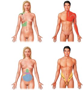
Pain can be classified as fast or slow, and sometimes is perceived in areas other than its origin (referred pain), due to visceral sensations carried by spinal nerves.
Mechanoreceptors: Tactile, Baroreceptors, and Proprioceptors
Mechanoreceptors are sensitive to stretch, compression, twisting, or distortion of cell membranes. They are divided into:
Tactile receptors: Detect touch, pressure, and vibration.
Baroreceptors: Monitor pressure changes in organs.
Proprioceptors: Monitor position of joints, muscle length, and tendon tension.
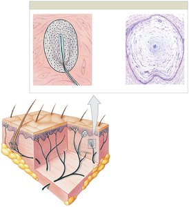
Tactile Receptors
Tactile receptors are further classified as unencapsulated or encapsulated:
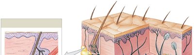
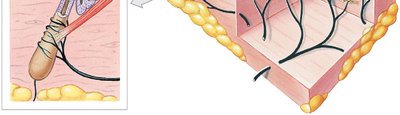
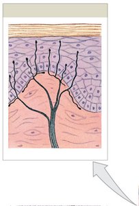
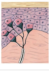
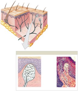
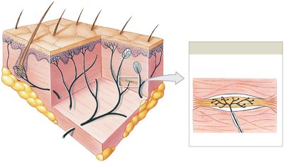
Unencapsulated: Free nerve endings, root hair plexus, tactile discs (Merkel cells).
Encapsulated: Tactile corpuscles (Meissner's), bulbous corpuscles (Ruffini), lamellar corpuscles (Pacinian).
Baroreceptors
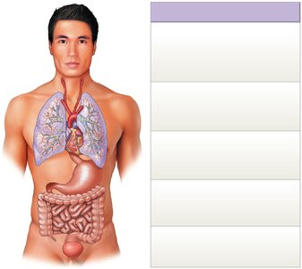
Baroreceptors provide information about pressure changes in various organs, contributing to autonomic regulation.
Located in carotid sinus, aortic sinus, lungs, digestive tract, colon, and bladder wall.
Trigger reflexes for cardiovascular, respiratory, digestive, and urinary functions.
Proprioceptors
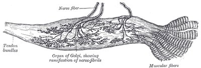
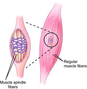
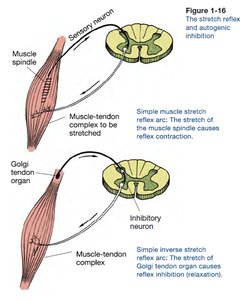
Proprioceptors monitor the position of joints, muscle length, and tension in tendons. Key types include:
Golgi tendon organs: Detect tension in tendons.
Muscle spindles: Detect muscle length and stretch.
Free nerve endings: Present in joint capsules.
Chemoreceptors
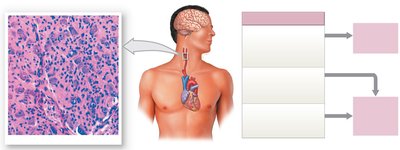
Chemoreceptors detect changes in concentrations of specific chemicals or compounds, such as pH, oxygen, and carbon dioxide. They are found in blood vessels and near respiratory centers.
Carotid bodies and aortic bodies monitor blood chemistry.
Medulla oblongata chemoreceptors monitor cerebrospinal fluid.
Special Senses: Vision
Overview of Vision
Vision is the most complex special sense, with receptors located in the eyes. The visual cortex is the largest of the special senses, responsible for processing detailed images.
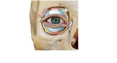
Accessory structures include eyelids, tarsal plates, tarsal glands, and the lacrimal apparatus.
Anatomy of the Eye
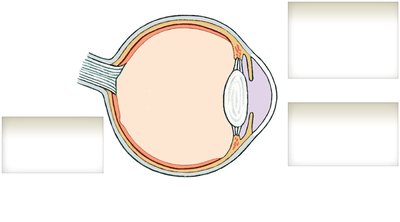
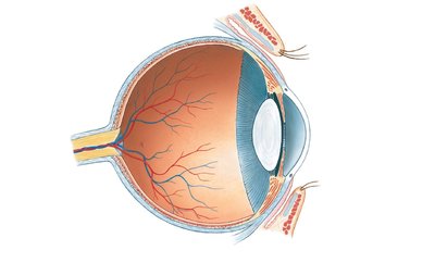
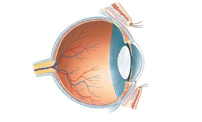
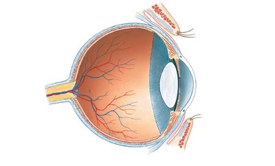
The wall of the eye consists of three layers:
Fibrous layer (outer): Sclera and cornea; provides support and protection, attachment for muscles, and helps focus vision.
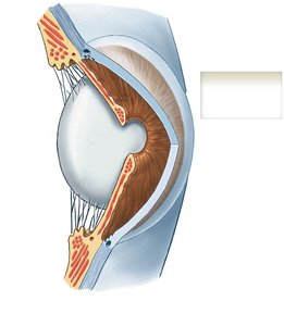
Vascular layer (intermediate): Iris, ciliary body, choroid; regulates light entry, secretes aqueous humor, controls lens shape.
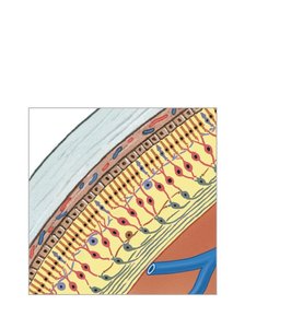
Inner layer (retina): Pigmented and neural layers; absorbs light, processes visual information.
Fibrous Layer
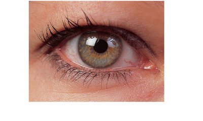
Sclera: White of the eye, insertion point for extra-ocular muscles.
Cornea: Transparent, avascular, allows light to enter.
Vascular Layer
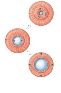
Iris: Contains smooth muscle, determines eye color, regulates pupil size.
Ciliary body: Holds lens in place, controls lens shape via ciliary zonule.
Choroid: Capillary network serving the retina.
Inner Layer (Retina)
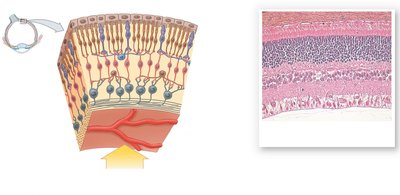
Pigmented layer: Absorbs light, interacts with photoreceptors.
Neural layer: Contains photoreceptors (rods and cones), supporting cells, neurons, and blood vessels.
Retina organization:
Photoreceptors: Rods (light sensitivity), cones (color vision).
Fovea centralis: Center of macula, highest visual acuity.
Bipolar cells: Synapse with rods and cones.
Ganglion cells: Generate action potentials, axons form optic nerve.
Optic disc: Blind spot where optic nerve exits.
Chambers and Fluids of the Eye
The eye contains three chambers:
Anterior chamber: Contains aqueous humor, provides nutrients and waste transport.
Posterior chamber: Also contains aqueous humor.
Vitreous chamber: Contains vitreous body, maintains eye shape and supports lens.
Lens and Accommodation
The lens focuses visual images on the retina by changing shape. Elastic fibers allow the lens to become more spherical for near vision and flatter for distant vision, controlled by the ciliary body and zonule.
Neural Pathways of Vision
Optic nerves (CN II) carry visual information to the brain. Fibers cross at the optic chiasm, travel to the lateral geniculate nucleus of the thalamus, and then to the visual cortex. Some fibers go to the superior colliculi (reflexive movements) and suprachiasmatic nucleus (circadian rhythms).
Clinical Anatomy: Macular Degeneration, Migraines, and Myopia
Macular Degeneration
Leading cause of blindness in older adults, involving deterioration of the macula (central vision area). No cure; vision loss progresses from central to peripheral.
Migraines
Migraine with aura: Flashes of light, blind spots, shimmering, affects both eyes.
Retinal migraine: Symptoms in one eye, temporary vision loss, possibly due to blood vessel constriction.
Myopia
Caused by axial elongation of the eyeball, resulting in nearsightedness (difficulty seeing distant objects).
Type | Description |
|---|---|
Macular Degeneration | Deterioration of macula, central vision loss |
Migraine with Aura | Visual disturbances, both eyes |
Retinal Migraine | Vision changes in one eye |
Myopia | Axial elongation, nearsightedness |
Additional info: The notes above expand on brief points from the original slides, providing definitions, examples, and context for each sensory system and clinical condition. Images included are directly relevant to the anatomical and physiological explanations provided.
