 뒤로
뒤로Study Guide: Joints (Articulations) in Human Anatomy & Physiology
스터디 가이드 - 스마트 노트
 자료에 맞춘 맞춤형 노트, 핵심 정의, 예시, 맥락을 확장해 제공합니다.
Joints (Articulations)
Introduction to Joints
Joints, also known as articulations, are the sites where two or more bones meet. They are essential for providing mobility to the skeleton and holding it together. Understanding the structure and function of joints is crucial for diagnosing and treating injuries such as sprains and dislocations.
Classification of Joints
Structural Classification
Joints are classified based on the material binding the bones and the presence or absence of a joint cavity. The three structural types are:
Fibrous Joints
Cartilaginous Joints
Synovial Joints
Functional Classification
Joints are also classified by the degree of movement they allow:
Synarthroses: Immovable joints
Amphiarthroses: Slightly movable joints
Diarthroses: Freely movable joints
Fibrous Joints
General Features
Fibrous joints are joined by dense fibrous connective tissue and lack a joint cavity. Most are immovable, though movement depends on the length of connective tissue fibers.
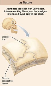
Sutures: Rigid, interlocking joints found only in the skull. Allow for growth during youth and ossify in adulthood to form synostoses.
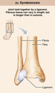
Syndesmoses: Bones connected by ligaments. Movement varies with fiber length; short fibers (e.g., inferior tibiofibular joint) allow little movement, while longer fibers (e.g., interosseous membrane between radius and ulna) allow more.
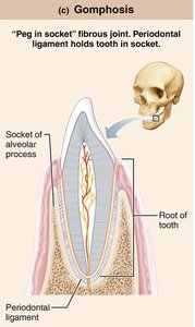
Gomphoses: Peg-in-socket joints, such as teeth in alveolar sockets, held by the periodontal ligament.
Cartilaginous Joints
General Features
Cartilaginous joints unite bones with cartilage and lack a joint cavity. They are not highly movable.
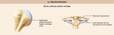
Synchondroses: Bones united by hyaline cartilage. Most are immovable (synarthrotic). Examples include the epiphyseal plate and the joint between the first rib and sternum.
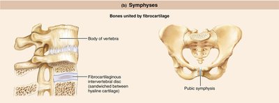
Symphyses: Bones united by fibrocartilage, with hyaline cartilage present as articular cartilage. These are strong, slightly movable (amphiarthrotic) joints, such as intervertebral joints and the pubic symphysis.
Synovial Joints
General Structure
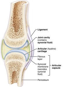
Synovial joints are characterized by a fluid-filled joint cavity and are all freely movable (diarthrotic). They include almost all limb joints and have six general features:
Articular cartilage: Hyaline cartilage covering bone ends, preventing crushing.
Joint (synovial) cavity: Unique, fluid-filled space.
Articular (joint) capsule: Two layers—external fibrous layer (dense irregular connective tissue) and inner synovial membrane (loose connective tissue producing synovial fluid).
Synovial fluid: Viscous, slippery filtrate lubricating and nourishing cartilage; contains phagocytic cells.
Reinforcing ligaments: Capsular, extracapsular, and intracapsular ligaments.
Nerves and blood vessels: Nerves detect pain and monitor position; capillary beds supply filtrate for synovial fluid.
Bursae and Tendon Sheaths
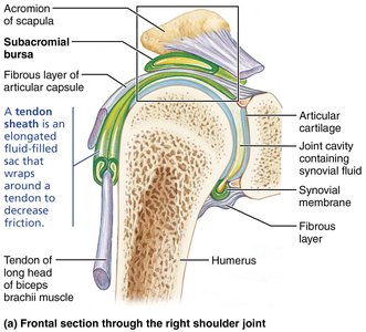
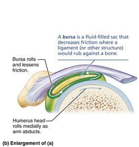
Bursae are fluid-filled sacs that reduce friction where ligaments, muscles, skin, tendons, or bones rub together. Tendon sheaths are elongated bursae wrapped around tendons subjected to friction.
Factors Influencing Stability
Shape of articular surfaces: Shallow surfaces are less stable than ball-and-socket.
Ligament number and location: More ligaments provide stronger support.
Muscle tone: Most important factor; keeps tendons taut and reinforces joints.
Movements Allowed by Synovial Joints
Types of Movement
Gliding: Flat bone surfaces slide across each other. Examples: intercarpal and intertarsal joints.
Angular Movements: Change the angle between bones. Includes flexion, extension, hyperextension, abduction, adduction, and circumduction.
Rotation: Bone turns around its own long axis (medial and lateral rotation).
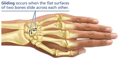
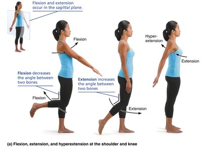
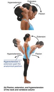
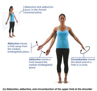
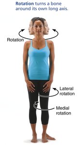
Table: Movements at Synovial Joints
Movement | Definition |
|---|---|
Gliding | Sliding the flat surfaces of two bones across each other |
Flexion | Decreasing the angle between two bones, usually in the sagittal plane |
Extension | Increasing the angle between two bones, usually in the sagittal plane |
Abduction | Moving a limb away from the body midline in the frontal plane |
Adduction | Moving a limb toward the body midline in the frontal plane |
Circumduction | Moving a limb or finger so that it describes a cone in space |
Rotation | Turning a bone around its longitudinal axis |
Medial rotation | Rotating toward the median plane |
Lateral rotation | Rotating away from the median plane |
Types of Synovial Joints
Six Types Based on Shape and Movement
Plane
Hinge
Pivot
Condylar (Ellipsoid)
Saddle
Ball-and-socket
Selected Synovial Joints
Temporomandibular Joint (TMJ)
The TMJ is a modified hinge joint where the mandibular condyle articulates with the temporal bone. It allows depression/elevation and side-to-side gliding. It is the most easily dislocated joint in the body.
Shoulder (Glenohumeral) Joint
The shoulder joint is a ball-and-socket joint, offering the greatest range of motion but sacrificing stability. The glenoid labrum deepens the cavity, and reinforcing ligaments and muscle tendons (especially the rotator cuff) provide stability.
Elbow Joint
The elbow is a hinge joint formed by the humerus, radius, and ulna. It allows flexion and extension only, with ligaments restricting side-to-side movement.
Hip (Coxal) Joint
The hip is a ball-and-socket joint with a deep acetabulum, providing stability and a good range of motion. Reinforcing ligaments and the acetabular labrum enhance stability.
Knee Joint
The knee is the largest and most complex joint, consisting of three joints surrounded by a single cavity. It includes the femoropatellar and tibiofemoral joints, stabilized by multiple ligaments and menisci.
Disorders of Joints
Common Joint Injuries
Cartilage tears: Often require arthroscopic surgery; meniscal transplants are possible.
Sprains: Ligaments are stretched or torn; healing is slow due to poor vascularization.
Dislocations (luxations): Bones are forced out of alignment; must be reduced to treat.
Inflammatory and Degenerative Conditions
Bursitis: Inflammation of bursae, treated with rest and anti-inflammatory drugs.
Tendonitis: Inflammation of tendon sheaths, usually due to overuse.
Arthritis: Over 100 types, including osteoarthritis (degenerative), rheumatoid arthritis (autoimmune), and gouty arthritis (uric acid crystal deposition).
Lyme disease: Caused by tick-borne bacteria, leading to joint pain and arthritis.
Developmental Aspects of Joints
By embryonic week 8, synovial joints resemble adult joints. Joint size, shape, and flexibility are modified by use. Aging leads to shortened ligaments and tendons, increased risk of disc herniation, and osteoarthritis. Full-range-of-motion exercise helps postpone joint problems.
Summary Table: Structural and Functional Characteristics of Body Joints
Joint Type | Binding Material | Joint Cavity | Movement | Examples |
|---|---|---|---|---|
Fibrous | Dense fibrous connective tissue | No | Immovable or slightly movable | Sutures, syndesmoses, gomphoses |
Cartilaginous | Cartilage | No | Immovable or slightly movable | Synchondroses, symphyses |
Synovial | Articular cartilage, synovial fluid | Yes | Freely movable | Shoulder, knee, hip, elbow |
