 뒤로
Study Guide: Selected Synovial Joints (Jaw, Shoulder, Elbow, Hip, Knee)
스터디 가이드 - 스마트 노트
 자료에 맞춘 맞춤형 노트, 핵심 정의, 예시, 맥락을 확장해 제공합니다.
Joints: Diversity of Synovial Joints
Overview of Synovial Joints
Synovial joints are the most movable and structurally complex type of joint in the human body. They share general features but also possess unique structural characteristics, abilities, and weaknesses. Five main synovial joints are commonly studied for their diversity: the temporomandibular (jaw), shoulder, elbow, hip, and knee joints.
Synovial Joint: A joint characterized by the presence of a synovial cavity, articular cartilage, and a joint capsule.
Key Features: Articulating bones, anatomical characteristics, movement allowed, and stability.
Temporomandibular Joint (TMJ)
Anatomical Features and Movements
The temporomandibular joint (TMJ), or jaw joint, is a modified hinge joint formed by the mandibular condyle articulating with the temporal bone. The posterior temporal bone forms the mandibular fossa, while the anterior portion forms the articular tubercle.
Articular Capsule: Thickens into a strong lateral ligament.
Movements:
Hinge: Depression and elevation of the mandible.
Gliding: Side-to-side (lateral excursion) grinding of teeth.
Stability: Most easily dislocated joint in the body due to its shallow socket.
Clinical Relevance: TMJ Dislocation
Dislocation is common, usually anterior, causing the mouth to remain open.
Symptoms: Ear and face pain, tender muscles, popping sounds, joint stiffness.
Causes: Grinding teeth, jaw trauma, poor occlusion.
Treatment: Bite plate, relaxing jaw muscles, manual realignment by a physician.


Shoulder (Glenohumeral) Joint
Anatomical Features and Stability
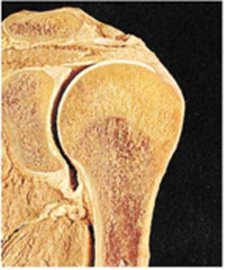
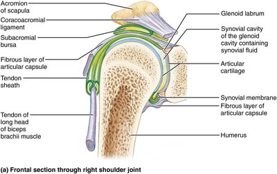
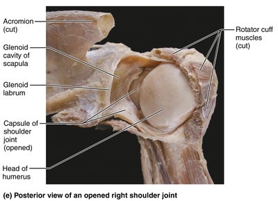
The shoulder joint is the most freely moving joint in the body, sacrificing stability for mobility. It is a ball-and-socket joint where the large, hemispherical head of the humerus fits into the small, shallow glenoid cavity of the scapula.
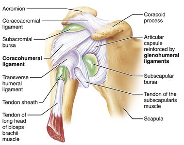
Articular Capsule: Thin and loose, contributing to freedom of movement.
Glenoid Labrum: Fibrocartilaginous rim that adds depth to the shallow cavity, but only holds one-third of the humeral head.
Reinforcing Ligaments:
Coracohumeral ligament: Supports weight of upper limb.
Three glenohumeral ligaments: Strengthen anterior capsule, but are weak supports.
Reinforcing Muscle Tendons:
Tendon of long head of biceps brachii: "Superstabilizer" securing humerus to glenoid cavity.
Four rotator cuff tendons: Subscapularis, Supraspinatus, Infraspinatus, Teres minor.
Clinical Relevance: Shoulder Dislocation
Dislocations are common due to mobility.
Weakest support is anterior and inferior; humerus can dislocate forward and downward.
Glenoid cavity provides poor support when humerus is rotated laterally and abducted.
Blows to top and back of shoulder can also cause dislocations.



Elbow Joint
Anatomical Features and Movements
The elbow joint is formed by the humerus articulating with the radius and ulna, consisting of three joints in one: humeroradial, humeroulnar, and proximal radioulnar joints. The hinge joint is primarily formed by the trochlear notch of the ulna articulating with the trochlea of the humerus.
Movement: Flexion and extension only.
Anular Ligament: Surrounds head of radius.
Capsular Ligaments:
Ulnar collateral ligament
Radial collateral ligament
Restrict side-to-side movement.

Hip (Coxal) Joint
Anatomical Features and Stability
The hip joint is a ball-and-socket joint where the large, spherical head of the femur articulates with the deep, cup-shaped acetabulum. It has a good range of motion, but is limited by the deep socket.
Acetabular Labrum: Rim of fibrocartilage that enhances depth of socket.
Reinforcing Ligaments:
Iliofemoral ligament
Pubofemoral ligament
Ischiofemoral ligament
Ligament of head of femur (ligamentum teres): Contains artery supplying head of femur, not important in stabilizing.
Stability: Greatest stability comes from deep ball-and-socket joint; hip dislocations are rare.

Knee Joint
Anatomical Features and Movements
The knee joint consists of three joints surrounded by a single cavity: the femoropatellar (patellofemoral) joint and the lateral and medial tibiofemoral joints. The femoropatellar joint is a plane joint allowing gliding motion during knee flexion, while the tibiofemoral joints are hinge joints allowing flexion, extension, and some rotation when the knee is partly flexed.
Joint Capsule: Thin and absent anteriorly.
Ligaments:
Medial and lateral patellar retinacula flank the patellar ligament.
Fibular and tibial collateral ligaments: Prevent rotation when knee is extended.
Oblique popliteal ligament: Stabilizes posterior knee joint.
Arcuate popliteal ligament: Reinforces joint capsule posteriorly.
Anterior cruciate ligament (ACL): Attaches to anterior tibia, prevents forward sliding of tibia and stops hyperextension.
Posterior cruciate ligament (PCL): Attaches to posterior tibia, prevents backward sliding of tibia and forward sliding of femur.
Bursae: At least 12 bursae associated with the knee joint.

Summary Table: Selected Synovial Joints
This table summarizes the main features of the five selected synovial joints.
Joint | Articulating Bones | Anatomical Features | Joint Type(s) | Movement Allowed | Stability |
|---|---|---|---|---|---|
Jaw (TMJ) | Mandible, Temporal bone | Mandibular condyle, articular capsule, lateral ligament | Modified hinge | Depression, elevation, lateral excursion | Low (easily dislocated) |
Shoulder | Humerus, Scapula | Glenoid cavity, labrum, ligaments, rotator cuff | Ball-and-socket | Flexion, extension, abduction, adduction, rotation | Moderate (sacrificed for mobility) |
Elbow | Humerus, Radius, Ulna | Trochlear notch, anular ligament, collateral ligaments | Hinge | Flexion, extension | High |
Hip | Femur, Pelvis | Acetabulum, labrum, ligaments | Ball-and-socket | Flexion, extension, abduction, adduction, rotation | High (deep socket) |
Knee | Femur, Tibia, Patella | Condyles, patellar ligament, collateral/cruciate ligaments | Hinge, plane | Flexion, extension, rotation (when flexed) | Moderate |
Additional info: Academic context was added to clarify anatomical features, clinical relevance, and movement types for each joint.
