 뒤로
Study Guide: Ch8C Selected Synovial Joints (Jaw, Shoulder, Elbow, Hip, Knee)
스터디 가이드 - 스마트 노트
 자료에 맞춘 맞춤형 노트, 핵심 정의, 예시, 맥락을 확장해 제공합니다.
Joints: Diversity and Structure of Synovial Joints
Overview of Synovial Joints
Synovial joints are the most movable and structurally complex type of joint in the human body. They share general features but also possess unique structural characteristics, abilities, and weaknesses. The five main synovial joints discussed here are the temporomandibular (jaw), shoulder, elbow, hip, and knee joints.
Definition: Synovial joints are characterized by the presence of a synovial cavity, articular cartilage, and a joint capsule.
Key Features: Allow a wide range of movements, are reinforced by ligaments and tendons, and are susceptible to injury due to their mobility.
Examples: Jaw, shoulder, elbow, hip, and knee joints.
Temporomandibular Joint (TMJ)
Anatomical Structure and Movements
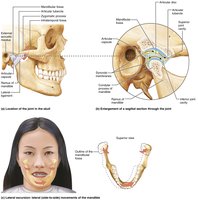
The temporomandibular joint (TMJ), or jaw joint, is a modified hinge joint formed by the articulation of the mandibular condyle with the temporal bone. It is the most easily dislocated joint in the body due to its shallow socket.
Articulating Bones: Mandibular condyle and temporal bone.
Anatomical Features: Mandibular fossa (posterior temporal bone), articular tubercle (anterior temporal bone), thickened articular capsule forming a strong lateral ligament.
Movements Allowed:
Hinge: Depression and elevation of the mandible.
Gliding: Side-to-side (lateral excursion) grinding movements.
Joint Stability: Most easily dislocated due to shallow socket.
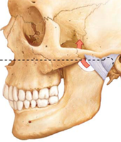
Clinical Relevance: TMJ Dislocation
Dislocation of the TMJ is common, usually occurring anteriorly and causing the mouth to remain open. Symptoms include ear and face pain, muscle tenderness, popping sounds, and joint stiffness. Causes include teeth grinding, trauma, or poor occlusion. Treatment may involve bite plates and muscle relaxation.
Shoulder (Glenohumeral) Joint
Anatomical Structure and Movements
The shoulder joint is the most freely moving joint in the body, sacrificing stability for mobility. It is a ball-and-socket joint formed by the large head of the humerus fitting into the shallow glenoid cavity of the scapula.
Articulating Bones: Humerus and scapula (glenoid cavity).
Anatomical Features: Glenoid labrum (fibrocartilaginous rim), thin and loose articular capsule, primarily anterior reinforcing ligaments (coracohumeral and glenohumeral ligaments).
Movements Allowed: Flexion, extension, abduction, adduction, rotation, circumduction.
Joint Stability: Stability is mainly provided by muscle tendons, especially the tendon of the long head of the biceps brachii and the four rotator cuff tendons (subscapularis, supraspinatus, infraspinatus, teres minor).

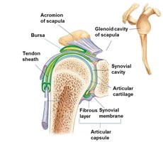
Clinical Relevance: Shoulder Dislocation
Shoulder dislocations are common due to the joint's mobility. The weakest points are anteriorly and inferiorly, allowing the humerus to dislocate forward and downward. Dislocations often occur during lateral rotation and abduction or from blows to the shoulder.
Elbow Joint
Anatomical Structure and Movements
The elbow joint consists of three joints in one: humeroradial, humeroulnar, and proximal radioulnar. The primary hinge joint is formed by the trochlear notch of the ulna articulating with the trochlea of the humerus, allowing flexion and extension only.
Articulating Bones: Humerus, radius, and ulna.
Anatomical Features: Anular ligament surrounds the head of the radius; ulnar and radial collateral ligaments restrict side-to-side movement.
Movements Allowed: Flexion and extension.
Joint Stability: Reinforced by strong ligaments.
Hip (Coxal) Joint
Anatomical Structure and Movements
The hip joint is a ball-and-socket joint formed by the large, spherical head of the femur articulating with the deep cup-shaped acetabulum of the pelvis. It allows a good range of motion but is limited by the deep socket, making dislocations rare.
Articulating Bones: Femur and pelvis (acetabulum).
Anatomical Features: Acetabular labrum (fibrocartilage rim), reinforcing ligaments (iliofemoral, pubofemoral, ischiofemoral, ligamentum teres).
Movements Allowed: Flexion, extension, abduction, adduction, rotation, circumduction.
Joint Stability: Greatest stability from deep ball-and-socket structure; ligamentum teres contains artery supplying femoral head.
Knee Joint
Anatomical Structure and Movements
The knee joint consists of three joints surrounded by a single cavity: femoropatellar (plane joint), lateral tibiofemoral, and medial tibiofemoral (hinge joints). It allows flexion, extension, and some rotation when partly flexed.
Articulating Bones: Femur, tibia, and patella.
Anatomical Features: Thin joint capsule, quadriceps tendon, patellar ligament, medial and lateral patellar retinacula, at least 12 bursae.
Movements Allowed: Flexion, extension, limited rotation.
Joint Stability: Stabilized by capsular, extracapsular, and intracapsular ligaments.
Fibular and tibial collateral ligaments prevent rotation when extended.
Oblique and arcuate popliteal ligaments stabilize posteriorly.
Anterior cruciate ligament (ACL) prevents forward sliding of tibia and hyperextension.
Posterior cruciate ligament (PCL) prevents backward sliding of tibia and forward sliding of femur.
Summary Table: Comparison of Selected Synovial Joints
This table summarizes the main features of the five selected synovial joints for quick review.
Joint | Articulating Bones | Type | Movements Allowed | Stability |
|---|---|---|---|---|
Temporomandibular (Jaw) | Mandible & Temporal Bone | Modified Hinge | Depression, Elevation, Lateral Excursion | Low (easily dislocated) |
Shoulder (Glenohumeral) | Humerus & Scapula | Ball-and-Socket | Flexion, Extension, Abduction, Rotation | Moderate (muscle tendons) |
Elbow | Humerus, Radius, Ulna | Hinge | Flexion, Extension | High (ligaments) |
Hip (Coxal) | Femur & Pelvis | Ball-and-Socket | Flexion, Extension, Abduction, Rotation | High (deep socket) |
Knee | Femur, Tibia, Patella | Hinge/Plane | Flexion, Extension, Rotation | Moderate (ligaments, tendons) |
Key Terms and Concepts
Synovial Joint: A freely movable joint with a synovial cavity.
Articular Capsule: The envelope surrounding a synovial joint.
Ligament: Connective tissue that stabilizes joints.
Labrum: Fibrocartilaginous rim that deepens a joint cavity.
Bursa: Fluid-filled sac reducing friction in joints.
Equations and Biomechanics
While synovial joint function is primarily anatomical, basic biomechanical principles apply:
Torque: (where is torque, is force, is lever arm distance)
Range of Motion: Determined by joint structure, ligament tension, and muscle tone.
Additional info:
Animations referenced in the original material provide dynamic views of joint movement and structure, useful for further study.
Clinical notes highlight common injuries and their anatomical basis, reinforcing the importance of joint structure in function and pathology.
