 뒤로
Study Guide: Spinal Cord, Spinal Nerves, and Reflexes
스터디 가이드 - 스마트 노트
 자료에 맞춘 맞춤형 노트, 핵심 정의, 예시, 맥락을 확장해 제공합니다.
Spinal Cord: Gross Anatomy
Overview and Structure
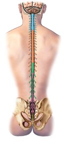
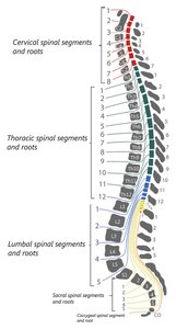
The spinal cord is a major component of the central nervous system, extending from the foramen magnum at the base of the skull to the inferior border of the L1 vertebra. It is responsible for transmitting neural signals between the brain and the rest of the body.
Conus medullaris: The tapered, terminal end of the spinal cord.
Filum terminale: A fibrous extension anchoring the spinal cord to the coccyx.
Cauda equina: A bundle of spinal nerve roots below the conus medullaris.
Regions: Cervical, thoracic, lumbar, and sacral.
Enlargements: Cervical and lumbosacral regions where nerves for limbs originate.
Spinal Cord: Sectional Anatomy
Gray and White Matter
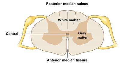
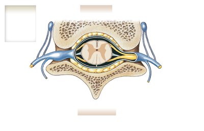
Sectional anatomy reveals a central H-shaped mass of gray matter surrounded by white matter. The gray matter contains neuron cell bodies and glial cells, while the white matter consists of myelinated axons organized into columns.
Gray matter: Organized into nuclei; posterior horns (sensory), anterior horns (motor), lateral horns (visceral motor).
White matter: Organized into columns (funiculi) containing tracts; ascending (sensory) and descending (motor).
Central canal: Runs through the center, containing cerebrospinal fluid.
Spinal Meninges
Protective Membranes
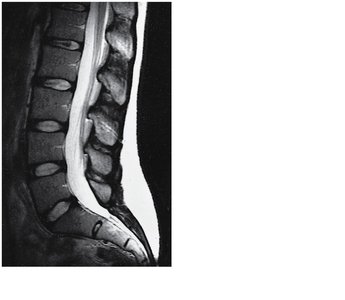
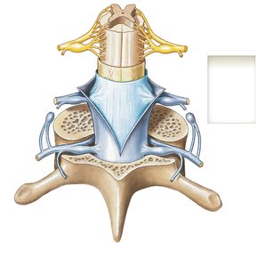
The spinal cord is protected by three specialized membranes called meninges, which provide stability and shock absorption. These layers are continuous with the cranial meninges and peripheral nerve connective tissue.
Dura mater: Tough, fibrous outer layer; continuous with epineurium.
Arachnoid mater: Middle layer; separated from pia mater by subarachnoid space containing cerebrospinal fluid.
Pia mater: Delicate inner layer; contains blood vessels and forms denticulate ligaments.
Spinal Nerves
Structure and Distribution
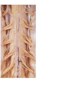
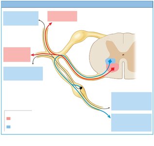
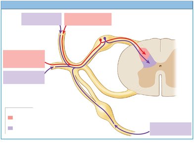
There are 31 pairs of spinal nerves, each formed by the fusion of dorsal (sensory) and ventral (motor) roots. Spinal nerves are mixed, containing both sensory and motor fibers, and are named according to their associated vertebrae.
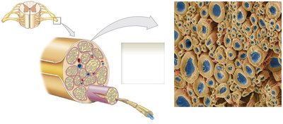
Connective tissue layers: Epineurium (outer), perineurium (middle, divides into fascicles), endoneurium (inner, surrounds individual axons).
Peripheral distribution: After fusion, nerves branch into dorsal ramus (serves neck/back) and ventral ramus (serves limbs/body wall). T1–L2 have additional rami communicantes for autonomic functions.
Dermatomes and Myotomes
Regional Innervation
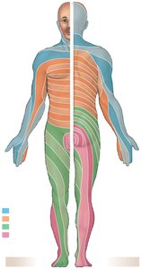
Dermatomes are regions of skin innervated by specific spinal nerves, while myotomes are muscle groups innervated by these nerves. This mapping is clinically important for diagnosing nerve injuries.
Dermatome: Area of skin supplied by a single spinal nerve.
Myotome: Group of muscles supplied by a single spinal nerve.
Nerve Plexuses
Cervical, Brachial, Lumbar, and Sacral Plexuses
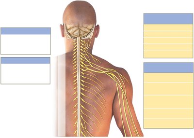
Nerve plexuses are networks of intersecting nerves that control skeletal muscles of the neck and limbs. The ventral rami blend fibers to form compound nerves, providing both sensory and motor innervation.
Cervical plexus (C1–C5): Innervates neck muscles and diaphragm.
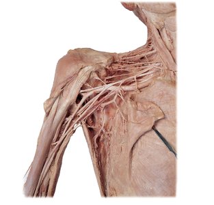
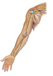
Brachial plexus (C5–T1): Innervates pectoral girdles and upper limbs.
Lumbar plexus (T12–L4): Innervates pelvic girdle and lower limbs.
Sacral plexus (L4–S4): Innervates lower limbs.
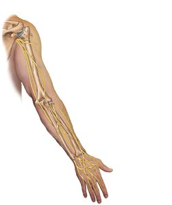
Brachial Plexus: Organization
Roots, Trunks, Divisions, Cords, and Terminal Nerves
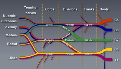
The brachial plexus is organized into roots, trunks, divisions, cords, and terminal nerves. This complex structure allows for extensive innervation of the upper limb.
Roots: Ventral rami of C5–T1.
Trunks: Superior (C5–C6), middle (C7), inferior (C8–T1).
Divisions: Each trunk splits into anterior and posterior divisions.
Cords: Lateral, posterior, and medial cords formed by divisions.
Terminal nerves: Musculocutaneous, axillary, median, radial, ulnar.
Reflexes
Reflex Arc and Types
Reflexes are immediate, involuntary motor responses to specific stimuli, crucial for maintaining homeostasis. The reflex arc is the neural pathway involved in a reflex action.
Reflex arc: Begins at a receptor, ends at a peripheral effector.
Monosynaptic reflex: Sensory neuron synapses directly onto motor neuron (e.g., stretch reflex).
Polysynaptic reflex: Involves interneurons between sensory and motor neurons, allowing for more complex responses.
Clinical Anatomy
Peripheral Nerve Palsies
Peripheral nerve palsies are regional losses of sensory and motor function due to nerve trauma or compression. Examples include brachial plexus birth palsy, pressure palsies, carpal tunnel syndrome, and sciatica.
Brachial plexus birth palsy: Injury to upper ventral rami of brachial plexus.
Pressure palsies: Temporary numbness or "pins and needles" sensation.
Carpal tunnel syndrome: Compression of the median nerve at the wrist.
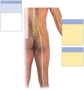
Sciatica: Compression or irritation of the sciatic nerve.
Lumbar Puncture and Epidural Block
These are clinical procedures involving the spinal cord and meninges. A lumbar puncture samples cerebrospinal fluid for diagnostic purposes, while an epidural block injects anesthetic into the epidural space to block sensory function.
Lumbar puncture: Needle inserted into subarachnoid space between L2 and sacrum.
Epidural block: Anesthetic injected into epidural space, affecting only local spinal nerves.
Summary Table: Major Nerves of the Lower Limb
Feature | Femoral Nerve | Sciatic Nerve |
|---|---|---|
Primary Region | Front of the lower body (Thigh and inner leg) | Back and lower portions of the leg (Thigh, calf, and foot) |
Motor Action | Straightens the knee, flexes the hip | Bends the knee, moves the ankle and toes |
Sensory Area | Front/inner thigh, inner calf down to the ankle | Back of thigh, entire outer calf, top and sole of the foot |
Additional info: The notes above expand on brief points from the original slides, providing definitions, examples, and clinical context for each topic. Images included are directly relevant to the anatomical structures and concepts discussed.
