 뒤로
뒤로Study Guide: The Appendicular Skeleton (Chapter 8)
스터디 가이드 - 스마트 노트
 자료에 맞춘 맞춤형 노트, 핵심 정의, 예시, 맥락을 확장해 제공합니다.
The Appendicular Skeleton
Overview
The appendicular skeleton consists of the bones that attach the limbs to the trunk and those of the limbs themselves. It is divided into the pectoral girdle (shoulder), pelvic girdle (hip), and the bones of the upper and lower limbs.
Pectoral girdle: Attaches the upper limbs to the trunk.
Pelvic girdle: Attaches the lower limbs to the trunk.
Upper and lower limbs differ in function but share a similar structural plan.
The Pectoral Girdle
Structure and Function
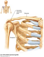
The pectoral girdle is composed of the clavicle and scapula. It provides attachment for muscles that move the upper limb and allows for a high degree of mobility.
Clavicle: Articulates medially with the manubrium and first rib, laterally with the scapula.
Scapula: Does not join each other or the axial skeleton, allowing free movement.
Only the clavicle articulates with the axial skeleton.
The socket of the shoulder joint (glenoid cavity) is shallow, favoring flexibility over stability.
Clavicle
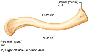
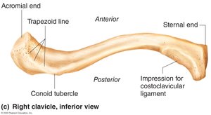
The clavicle extends horizontally across the superior thorax and serves as a strut to keep the scapula and arms laterally positioned.
Sternal end: Articulates with the manubrium.
Acromial end: Articulates with the scapula.
Transmits compression forces from the upper limbs to the axial skeleton.
Provides attachment for muscles.
Scapula
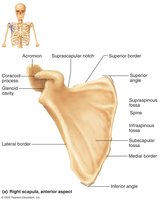
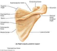
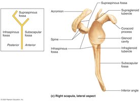
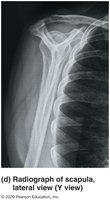
The scapula lies on the dorsal surface of the rib cage, between ribs 2–7. It has three borders (superior, medial, lateral) and three angles (lateral, superior, inferior).
Provides attachment for muscles.
Allows for a wide range of motion of the upper limb.
The Upper Limb
Bone Groups
Each upper limb contains thirty bones, grouped into three sets:
Arm: Humerus
Forearm: Radius and ulna
Hand: Carpals, metacarpals, phalanges
Arm (Humerus)
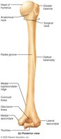
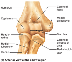
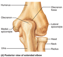
The humerus is the only bone of the arm, articulating with the scapula at the shoulder and with the radius and ulna at the elbow. It is the longest and strongest bone of the upper limb. 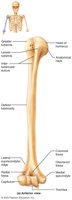
Forearm
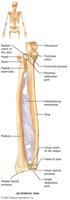
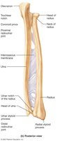
The forearm is formed from the radius and ulna.
Proximal ends articulate with the humerus; distal ends with the carpals.
Radius and ulna articulate with each other at the proximal and distal radioulnar joints.
The interosseous membrane interconnects the radius and ulna.
In anatomical position, the radius is lateral and the ulna is medial.
Ulna
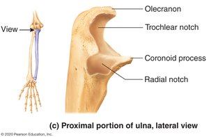
Main bone responsible for forming the elbow joint with the humerus.
Hinge joint allows forearm to bend on arm.
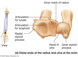
Distal end separated from carpals by fibrocartilage; plays little role in hand movement.
Major landmarks: Olecranon, radial notch, trochlear notch, coronoid process, ulnar styloid process.
Radius
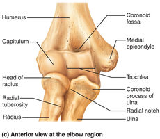
Superior surface of the head articulates with the capitulum.
Medially, head articulates with the radial notch of the ulna.
Contributes heavily to the wrist joint; distal radius articulates with carpal bones.
Major landmarks: Head, neck, radial tuberosity, radial styloid process.
Hand
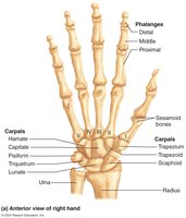
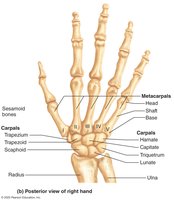
The hand includes the carpals (wrist), metacarpals (palm), and phalanges (fingers).
Carpus
Forms the true wrist; composed of eight marble-sized bones arranged in two rows.
Proximal row (lateral to medial): Scaphoid, lunate, triquetrum, pisiform.
Distal row (lateral to medial): Trapezium, trapezoid, capitate, hamate.
Mnemonic: "Sally left the party to take Carmen home".
Metacarpus
Five metacarpals radiate distally from the wrist, forming the palm.
Numbered beginning with the pollex (thumb).
Bases articulate proximally with carpals; heads articulate distally with proximal phalanges.
Phalanges
Numbered beginning with the pollex (thumb).
Each finger (except thumb) has three phalanges: proximal, middle, distal.
The Pelvic Girdle
Structure and Function
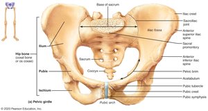
The pelvic girdle attaches the lower limbs to the spine and supports visceral organs. It is more stable than the pectoral girdle, with less freedom of movement.
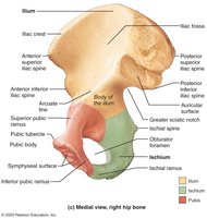
Consists of paired hip bones (coxal bones) and the sacrum.
Coxal bones unite anteriorly and articulate posteriorly with the sacrum.
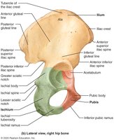
In childhood, consists of three separate bones: ilium, ischium, pubis.
Acetabulum: Deep socket on lateral pelvic surface, receives the head of the femur.
Ilium
Large, flaring bone forming the superior region of the coxal bone.
Site of attachment for many muscles.
Articulates with the sacrum at the sacroiliac joint.
Ischium
Forms posteroinferior region of the coxal bone.
Joins the pubis anteriorly.
Ischial tuberosities are the strongest part of the hip bone.
Pubis
Forms the anterior region of the coxal bone.
Pubic symphysis: Two pubic bones joined by fibrocartilage at the midline.
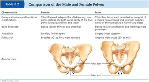
Pubic arch: Angle helps distinguish male from female pelvis.
Pelvic Structure and Childbearing
The bony pelvis is divided into two regions:
False (greater) pelvis: Bounded by alae of the iliac bones.
True (lesser) pelvis: Inferior to pelvic brim, forms a bowl containing pelvic organs.
Comparison of Male and Female Pelves
Characteristic | Female | Male |
|---|---|---|
General structure and functional modifications | Tilted forward, adapted for childbearing; true pelvis defines birth canal; cavity of true pelvis is broad, shallow, and rounded | Tilted less forward, adapted for support of heavier build and stronger muscles; cavity of true pelvis is narrow and deep |
Bones thickness | Bones lighter, thinner, and smoother | Bones heavier and thicker, markings more prominent |
Acetabula | Smaller, farther apart | Larger, closer together |
Pubic arch | Broader (80°–90°), more rounded | Angle is more acute (50°–60°) |
The Lower Limb
Overview
The lower limb carries the entire weight of the erect body. Its bones are thicker and stronger than those of the upper limb. It is divided into three segments: thigh, leg, and foot.
Thigh
Region between hip and knee.
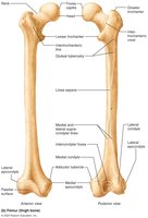
Femur: Single bone of the thigh; longest and strongest bone in the body.
Ball-shaped head articulates with the acetabulum.
Patella
Triangular sesamoid bone embedded in the tendon securing the quadriceps muscles.
Protects the knee anteriorly and improves leverage of thigh muscles across the knee.
Leg
Region between knee and ankle.
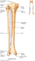
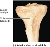
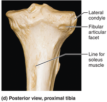
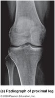
Tibia: More massive medial bone, receives weight from femur.
Fibula: Sticklike lateral bone, stabilizes the ankle joint.
Interosseous membrane connects tibia and fibula.
Foot
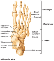
The foot is composed of the tarsus, metatarsus, and phalanges. It supports body weight and acts as a lever for walking. Segmentation makes the foot pliable and adapted to uneven ground.
Tarsus
Posterior half of the foot; contains seven tarsal bones.
Body weight is borne primarily by the talus and calcaneus.
Other tarsals: Cuboid, navicular, medial/intermediate/lateral cuneiforms.
Metatarsus
Five small long bones called metatarsals, numbered beginning with the hallux (great toe).
First metatarsal supports body weight.
Phalanges of the Toes
Fourteen phalanges; smaller and less nimble than fingers.
Each toe has proximal, middle, and distal phalanges, except the great toe (hallux), which has only two.
Summary Table: Bones of the Appendicular Skeleton
Region | Main Bones | Key Functions |
|---|---|---|
Pectoral Girdle | Clavicle, Scapula | Attaches upper limb, mobility |
Upper Limb | Humerus, Radius, Ulna, Carpals, Metacarpals, Phalanges | Manipulation, movement |
Pelvic Girdle | Ilium, Ischium, Pubis, Sacrum | Attaches lower limb, stability |
Lower Limb | Femur, Tibia, Fibula, Tarsals, Metatarsals, Phalanges | Support, locomotion |
Key Terms and Definitions
Girdle: Structure that attaches limbs to the trunk.
Acetabulum: Socket in the pelvic bone for the femur.
Interosseous membrane: Fibrous sheet connecting two bones (radius/ulna, tibia/fibula).
Sesamoid bone: Bone embedded within a tendon (e.g., patella).
Additional info:
The appendicular skeleton is essential for movement and manipulation of the environment, as well as for supporting the body during locomotion.
Differences in male and female pelvises are important for childbirth and can be used in forensic identification.
