 뒤로
Study Guide: The Axial and Appendicular Skeleton
스터디 가이드 - 스마트 노트
 자료에 맞춘 맞춤형 노트, 핵심 정의, 예시, 맥락을 확장해 제공합니다.
The Axial Skeleton
Overview of Skull Structure
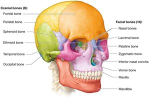
The skull is a complex structure composed of 22 bones, organized into cranial and facial groups. Cranial bones protect the brain, while facial bones form the framework of the face and support sensory organs.
Cranial bones (8): Frontal, parietal (paired), sphenoid, ethmoid, temporal (paired), occipital
Facial bones (14): Nasal (paired), lacrimal (paired), palatine (paired), zygomatic (paired), inferior nasal concha (paired), maxilla (paired), mandible, vomer
Sutures: Immovable joints connecting skull bones, except the mandible
Skull Cavities and Sinuses
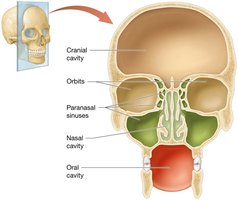
The skull contains several cavities that house sensory organs and air-filled spaces called sinuses. These structures are essential for functions such as smell, taste, vision, hearing, and balance.
Paranasal sinuses: Air-filled spaces in four bones surrounding the nasal cavity
Orbits: Contain the eyeballs
Nasal cavity: Houses olfactory receptors
Oral cavity: Surrounds teeth and tongue
Cranial cavity: Encloses the brain
The Fetal Skull
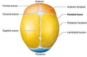
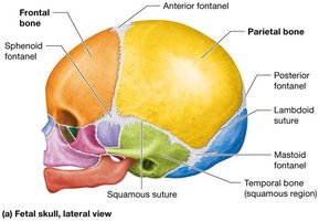
Unlike the adult skull, the fetal skull bones are not fused, allowing flexibility during birth and growth. Membranous areas called fontanels are present until the cranial bones complete development.
Fontanels: Soft spots between fetal cranial bones
Sutures: Unfused in infants, allowing skull expansion
Development: Fontanels close by 18–24 months
Hyoid Bone
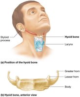
The hyoid bone is unique because it does not articulate with any other bone. It is suspended by ligaments and serves as an attachment point for muscles involved in swallowing and speech.
Location: Near the larynx, attached to temporal bone by ligaments
Function: Supports tongue and muscles of the neck
The Vertebral Column and Thoracic Cage
Overview of the Vertebral Column
The vertebral column, or spine, consists of 33 vertebrae classified by region and function. It provides structural support, protects the spinal cord, and allows flexibility.
Cervical (C1–C7): Neck region
Thoracic (T1–T12): Articulate with ribs
Lumbar (L1–L5): Lower back, weight-bearing
Sacral (5 fused): Form sacrum, articulate with pelvis
Coccygeal (3–5 fused): Form coccyx, most inferior
Structure of the Vertebrae
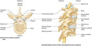
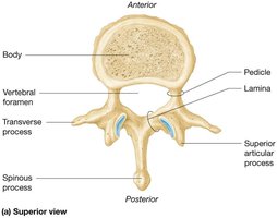
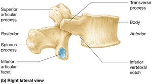
All vertebrae share common structural features that allow for movement and protection of the spinal cord.
Body: Main weight-bearing portion
Vertebral foramen: Passage for spinal cord
Pedicles and laminae: Form vertebral arch
Articular processes: Allow articulation between vertebrae
Transverse and spinous processes: Sites for muscle attachment
Structure of the Cervical Vertebrae
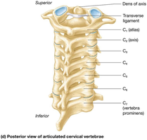
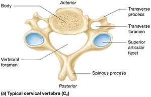
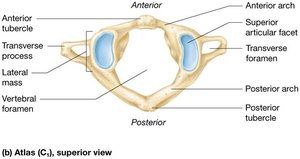
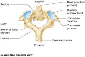
Cervical vertebrae are the smallest and are easily identified by the presence of transverse foramina. The first two cervical vertebrae, atlas (C1) and axis (C2), have unique structures.
Transverse foramina: Allow passage of vertebral arteries
C1 (Atlas): Lacks a body, has anterior and posterior arches
C2 (Axis): Contains the dens (odontoid process) for head rotation
Structure of the Thoracic Vertebrae
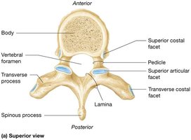
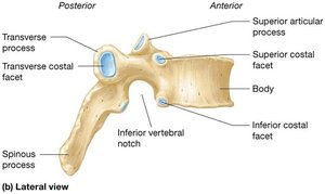
Thoracic vertebrae are larger than cervical vertebrae and have features for rib articulation. Their spinous processes point inferiorly.
Costal facets: Articulate with ribs
Heart-shaped bodies: Distinctive shape
Long spinous processes: Point downward
Structure of the Lumbar Vertebrae
Lumbar vertebrae are the largest and heaviest, designed to bear the weight of the torso. They have thick bodies and short spinous processes.
Kidney-shaped bodies: Adapted for weight-bearing
Flattened vertebral foramina: Triangular shape
Thick pedicles and laminae: Provide strength
Structure of the Sacrum
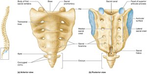
The sacrum consists of five fused vertebrae forming the posterior boundary of the pelvic cavity. It contains openings for nerve passage and articulates with the pelvis and coccyx.
Base: Articulates with L5
Apex: Articulates with coccyx
Sacral canal: Continuation of vertebral canal
Sacral foramina: Passage for nerves
Structure of the Coccyx
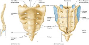
The coccyx is formed by three to five fused vertebrae and represents the terminal end of the vertebral column. It provides attachment for ligaments and muscles.
Coccygeal cornu: Hornlike projections
Intervertebral Discs
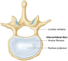
Intervertebral discs are fibrocartilage pads between vertebrae that absorb shock and bind the column together. Each disc has a soft nucleus pulposus and a tough anulus fibrosus.
Nucleus pulposus: Jelly-like center, shock absorber
Anulus fibrosus: Outer ring, binds vertebrae
The Thoracic Cage
Structure of the Thoracic Cage
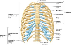
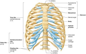
The thoracic cage consists of the sternum, ribs, and thoracic vertebrae. It protects vital organs and provides attachment points for muscles involved in respiration.
Sternum: Composed of manubrium, body, and xiphoid process
Ribs: 12 pairs, classified as true (1–7), false (8–12), and floating (11–12)
Costal cartilages: Connect ribs to sternum
The Appendicular Skeleton
The Pectoral Girdle
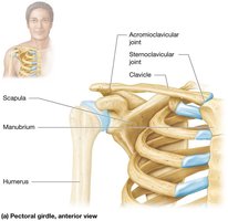
The pectoral girdle consists of the clavicle and scapula, which connect the upper limb to the axial skeleton and allow for a wide range of motion.
Clavicle: Articulates with sternum and scapula
Scapula: Provides attachment for muscles and articulates with humerus
Clavicle
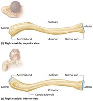
The clavicle, or collarbone, is a slender bone that acts as a strut between the sternum and scapula. It is important for shoulder stability.
Sternal end: Medial, articulates with manubrium
Acromial end: Lateral, articulates with scapula
Scapula
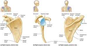
The scapula is a flat, triangular bone with several important features for muscle attachment and articulation with the humerus.
Acromion: Articulates with clavicle
Coracoid process: Muscle attachment
Glenoid cavity: Articulates with humerus
Spine: Prominent ridge
Bones of the Forearm
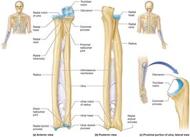
The forearm consists of the radius and ulna, which are connected by an interosseous membrane. Both bones articulate with the humerus and carpal bones.
Radius: Lateral bone
Ulna: Medial bone
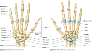
Bones of the Wrist: Carpals
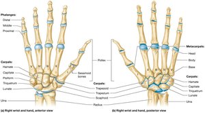
The wrist contains eight carpal bones arranged in two rows. These bones allow for flexibility and movement of the hand.
Proximal row: Scaphoid, lunate, triquetrum, pisiform
Distal row: Trapezium, trapezoid, capitate, hamate
Bones of the Hand and Fingers: Metacarpals and Phalanges
The hand consists of five metacarpals and fourteen phalanges. Each finger has three phalanges, except the thumb, which has two.
Metacarpals: Numbered I–V from lateral to medial
Phalanges: Proximal, middle, distal (thumb lacks middle)
Bones of the Pelvic Girdle and Lower Limb
Pelvic Girdle Structure
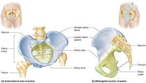
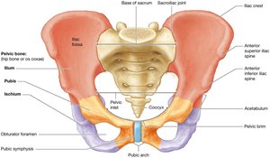
The pelvic girdle is formed by two coxal bones and the sacrum. It supports the weight of the upper body and protects pelvic organs.
Coxal bones: Each consists of ilium, ischium, and pubis
Acetabulum: Deep socket for femur articulation
Pelvic Differences Between Genders
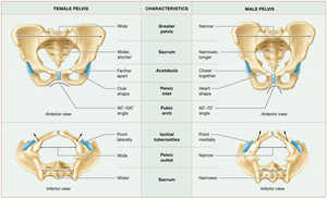
The pelvis differs between males and females, with the female pelvis adapted for childbirth. Key differences include width, shape, and angle of the pubic arch.
Characteristic | Female Pelvis | Male Pelvis |
|---|---|---|
Greater pelvis | Wide | Narrow |
Acetabula | Farther apart | Closer together |
Pubic arch | 60–100° | 60–70° |
Sacrum | Wide | Narrow |
Pelvic outlet | Wider | Narrower |
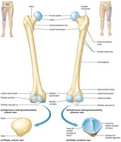
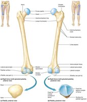
The Femur and Patella
The femur is the largest bone in the body, articulating with the pelvis at the hip joint and with the tibia at the knee. The patella is a sesamoid bone within the tendon of the thigh muscle.
Femur: Head, greater and lesser trochanters, epicondyles, condyles
Patella: Triangular, articulates with femur
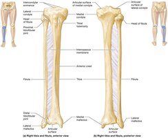
Bones of the Leg: Tibia and Fibula
The tibia and fibula are the bones of the leg, connected by an interosseous membrane. The tibia bears weight, while the fibula provides stability.
Tibia: Medial, weight-bearing
Fibula: Lateral, slender
Bones of Ankle and Foot: Tarsals, Metatarsals, and Phalanges
The ankle and foot contain seven tarsal bones, five metatarsals, and fourteen phalanges. The talus articulates with the leg bones, and the calcaneus forms the heel.
Proximal tarsals: Talus, calcaneus, navicular
Distal tarsals: Medial, intermediate, lateral cuneiform, cuboid
Metatarsals: Numbered I–V
Phalanges: Great toe (hallux) has two, others have three
Study Boost: Remembering Skull Bones and Vertebrae
Mnemonic Devices
Mnemonics are useful for memorizing the names and numbers of bones in the skull and vertebral column.
Pest Of 6: Parietal, Ethmoid, Sphenoid, Temporal, Occipital, Frontal (cranial bones)
Virgil Is Now Making My Pet Zebra Laugh: Vomer, Inferior nasal conchae, Nasal, Mandible, Maxillae, Palatine, Zygomatic, Lacrimal (facial bones)
Breakfast at 7, lunch at 12, dinner at 5: 7 cervical, 12 thoracic, 5 lumbar vertebrae
Visual Analogies
Sphenoid bone: Resembles a bat with wings
Ethmoid bone: Like an iceberg, only tip visible
Thoracic vertebra: Looks like a giraffe's head
Lumbar vertebra: Resembles a moose's head
Lab Activities
Studying the bones of the skull
Studying the bones of the vertebral column and thoracic cage
Identifying bones in a bag
Studying the bones of the appendicular skeleton
Compare the Vertebrae
Comparing cervical, thoracic, and lumbar vertebrae helps in identification and understanding their functional adaptations.
Cervical: Small, transverse foramina
Thoracic: Heart-shaped body, costal facets
Lumbar: Large, kidney-shaped body
Summary Table: Vertebral Column Regions
Region | Number of Vertebrae | Key Features |
|---|---|---|
Cervical | 7 | Transverse foramina, small size |
Thoracic | 12 | Costal facets, heart-shaped body |
Lumbar | 5 | Large, kidney-shaped body |
Sacral | 5 (fused) | Forms sacrum |
Coccygeal | 3–5 (fused) | Forms coccyx |
