 뒤로
뒤로Study Guide: The Axial Skeleton in Human Anatomy & Physiology
스터디 가이드 - 스마트 노트
 자료에 맞춘 맞춤형 노트, 핵심 정의, 예시, 맥락을 확장해 제공합니다.
The Axial Skeleton
Overview of the Axial Skeleton
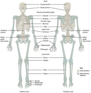
The axial skeleton forms the central axis of the human body and consists of 80 bones, accounting for approximately 40% of the total bones. It includes the skull, vertebral column, and thoracic cage. The axial skeleton provides structural support, protects vital organs, and serves as attachment points for muscles.
Skull: 22 bones (8 cranial, 14 facial)
Bones associated with the skull: 6 auditory ossicles, 1 hyoid bone
Thoracic cage: Sternum and 24 ribs
Vertebral column: 24 vertebrae, sacrum, coccyx
The Skull
Structure and Function
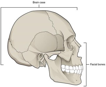
The skull is composed of the cranium (brain case) and facial bones. The cranium encases and protects the brain, while the facial bones form the structure of the face, including the jaws, nasal cavities, and orbits.
Cranial bones: 8 bones forming the brain case
Facial bones: 14 bones forming the face
Cranial Bones
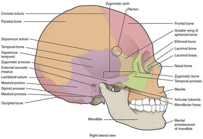
The cranial bones are paired and unpaired, forming the upper lateral, lower lateral, anterior, and posterior regions of the skull.
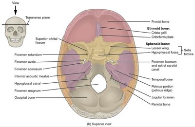
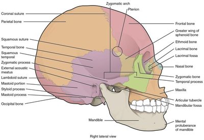
Parietal bones: Form most of the upper lateral side of the skull; joined at the top, bounded by frontal, temporal, and occipital bones.
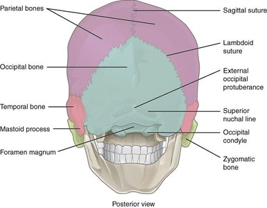
Temporal bones: Form the lower lateral side; include the mastoid process (muscle attachment for head rotation).
Frontal bone: Forms the forehead and floor of the anterior cranial cavity.
Occipital bone: Forms the posterior skull and base; contains the foramen magnum (spinal cord exit) and occipital condyles (articulation with C1).
Sphenoid bone: Forms part of the cranial floor, acts as a cross-bridge, unites cranial and facial bones, contains optic canals and sella turcica (pituitary gland housing).
Ethmoid bone: Forms part of the anterior cranial floor, roof of the nasal cavity, part of the nasal septum, and medial orbit wall; includes cribriform plate (olfactory foramina), perpendicular plate, crista galli, and ethmoidal labyrinth.
Sutures of the Skull
Sutures are immobile joints between adjacent skull bones, providing stability and protection.
Coronal suture: Attaches frontal bone to parietal bones
Sagittal suture: Attaches right and left parietal bones
Lambdoid suture: Attaches occipital bone to parietal and temporal bones
Squamous suture: Attaches temporal bone to parietal bone
Facial Bones
The facial bones form the structure of the face, including the upper and lower jaws, nasal cavity, and orbits.
Maxillary bones: Form the upper jaw, most of the hard palate, medial floor of the orbit, and lateral base of the nose
Palatine bones: Form the posterior portion of the hard palate
Zygomatic bones: Cheekbones; form lateral wall and margin of the orbit
Nasal bones: Form the bridge of the nose
Lacrimal bones: Form the anterior, medial wall of the orbit
Inferior nasal conchae: Thin, curved plates projecting into the nasal cavity
Vomer: Forms the posterior-inferior part of the nasal septum
Mandible: Forms the lower jaw; only moveable skull bone
Mandible Structure
The mandible consists of a horizontal body and a vertically oriented ramus. It features the coronoid process (muscle attachment), condylar process (articulates with temporal bone), alveolar process (anchors lower teeth), mental protuberance (chin), mental foramen (nerve passage), and mandibular foramen (blood vessel and nerve passage).
The Orbit and Nasal Structures
The orbit is a bony socket housing the eyeball, formed by seven skull bones. The nasal septum is composed of bone and cartilage, including the perpendicular plate of the ethmoid bone and the vomer. Nasal conchae create air turbulence, promoting warming and humidification of inhaled air.
Cranial Fossae
The floor of the cranial cavity is divided into three fossae:
Anterior cranial fossa: Contains frontal lobes
Middle cranial fossa: Contains temporal lobes; includes optic canal, foramen rotundum, foramen ovale
Posterior cranial fossa: Contains cerebellum; includes internal acoustic meatus, hypoglossal canal, jugular foramen
Paranasal Sinuses
Paranasal sinuses are air-filled spaces within certain skull bones (frontal, sphenoid, ethmoid, maxillary). They are lined with mucous epithelium, connected to the nasal cavities, and help warm, humidify, and filter inhaled air.
Hyoid Bone
The hyoid bone is a U-shaped bone in the upper neck, not connected to any other bone. It serves as an attachment site for muscles of the larynx, pharynx, and tongue.
The Vertebral Column
Structure and Regions
The vertebral column, or spine, is composed of 26 bones: 24 vertebrae, the sacrum, and the coccyx. It provides structural support, protects the spinal cord, and allows flexibility.
Cervical vertebrae (C1–C7): Located in the neck; C1 (atlas) articulates with occipital condyles, C2 (axis) allows head rotation
Thoracic vertebrae (T1–T12): Located in the upper back; articulate with ribs
Lumbar vertebrae (L1–L5): Located in the lower back; largest and bear most weight
Sacrum: Fusion of 5 vertebrae; part of the pelvis
Coccyx: Fusion of 3–5 vertebrae; tailbone
General Vertebra Structure
Each vertebra consists of a vertebral body (anterior, weight-bearing), vertebral arch (posterior), pedicles (lateral sides), laminae (roof), vertebral foramen (spinal cord passage), intervertebral foramen (spinal nerve passage), transverse and spinous processes (muscle attachment), and articular processes (vertebral articulation).
Intervertebral Discs
Intervertebral discs are composed of fibrocartilage and are found between vertebrae. They act as shock absorbers and allow flexibility.
The Thoracic Cage and Ribs
Structure and Function
The thoracic cage consists of 12 pairs of ribs, costal cartilages, the sternum, and thoracic vertebrae. It protects vital organs and supports respiration.
Sternum: Composed of manubrium (superior, articulates with clavicles and 1st ribs), body (articulates with ribs 2–7), and xiphoid process (inferior, muscle attachment)
Ribs: Elongate, curved bones; posteriorly articulate with thoracic vertebrae
True ribs (vertebrosternal): Rib pairs 1–7, directly connect to sternum via costal cartilage
False ribs (vertebrochondral): Rib pairs 8–10, indirectly connect to sternum via costal cartilage of rib 7
Floating ribs (vertebral): Rib pairs 11–12, do not attach to sternum
Summary Table: Axial Skeleton Components
Region | Main Bones | Key Functions |
|---|---|---|
Skull | Cranial, Facial, Auditory ossicles, Hyoid | Protects brain, forms face, supports sensory organs |
Vertebral Column | Cervical, Thoracic, Lumbar, Sacrum, Coccyx | Supports body, protects spinal cord, allows movement |
Thoracic Cage | Sternum, Ribs, Thoracic vertebrae | Protects heart/lungs, supports respiration |
Key Terms and Definitions
Axial skeleton: Central skeleton including skull, vertebral column, and thoracic cage
Cranial bones: Bones forming the brain case
Facial bones: Bones forming the structure of the face
Suture: Immobile joint between skull bones
Foramen: Opening in bone for nerves and blood vessels
Fossa: Depression in bone
Process: Projection from bone for muscle attachment or articulation
Example: Clinical Relevance
Damage to the occipital bone or foramen magnum can affect the spinal cord. Fractures of the maxilla or mandible impact chewing and speech. Sinus infections can affect the paranasal sinuses, leading to pain and congestion.
Additional info: Academic context was added to clarify bone functions, clinical relevance, and structural relationships.
