 뒤로
Study Guide: The Brain and Cranial Nerves (Anatomy & Physiology)
스터디 가이드 - 스마트 노트
 자료에 맞춘 맞춤형 노트, 핵심 정의, 예시, 맥락을 확장해 제공합니다.
Ch. 16: The Brain and Cranial Nerves
Introduction to the Brain
The brain is the central organ of the nervous system, responsible for integrating sensory information, generating thoughts, memories, and behaviors. It contains approximately 20 billion neurons and constitutes 95% of the neural tissue in the body. The brain's consistency is similar to medium-firm tofu or chilled gelatin, highlighting its delicate nature.
Complexity: Integrates multiple signals simultaneously to adapt to changing circumstances.
Contrast: Unlike spinal reflexes, the brain processes higher-order functions.
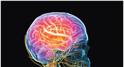
Major Divisions of the Brain
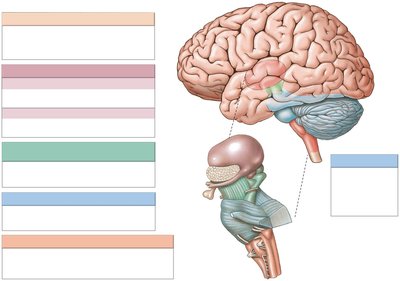
The brain is divided into six major regions, each with distinct functions and anatomical features. Understanding these divisions is essential for recognizing their roles in neural processing and clinical relevance.
Medulla Oblongata: Controls autonomic functions and relays sensory information.
Pons: Connects cerebellum to brainstem; involved in motor control.
Mesencephalon (Midbrain): Processes visual and auditory data; coordinates reflexive motor responses.
Diencephalon: Includes thalamus and hypothalamus; relays sensory information and controls emotions.
Cerebellum: Coordinates complex somatic motor patterns.
Cerebrum: Responsible for conscious thought, memory, and voluntary movement.
Gray Matter vs. White Matter
The brain's internal structure is organized into gray and white matter, each serving distinct functions:
Gray Matter: Composed of neuron cell bodies (nuclei); surrounds ventricles and passageways.
White Matter: Consists of axon tracts; in cerebrum and cerebellum, white matter is covered by a superficial layer of gray matter (cortex).
Protection and Support of the Brain
Cranial Meninges
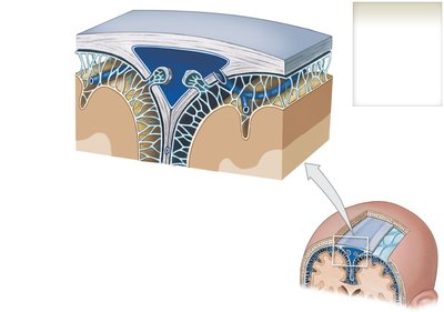
The brain is protected by three layers of cranial meninges, which are continuous with the spinal meninges. These layers provide structural support and help prevent injury.
Dura Mater: Outermost, tough layer; consists of periosteal and meningeal layers.
Arachnoid Mater: Middle, delicate membrane; covers the brain and contains the subarachnoid space.
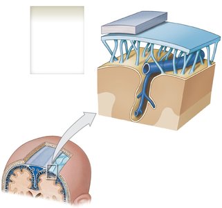
Pia Mater: Innermost, thin layer; tightly adheres to the brain's surface and is highly vascular.
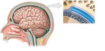
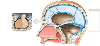
Dura Mater and Meningeal Folds
The dura mater forms folds that subdivide the cranial cavity and limit brain movement. These folds include:
Falx Cerebri: Separates cerebral hemispheres; contains sagittal sinuses.
Tentorium Cerebelli: Separates cerebrum from cerebellum; contains transverse sinus.
Falx Cerebelli: Divides cerebellar hemispheres.
Diaphragma Sellae: Anchors dura mater to sphenoid bone; surrounds pituitary gland.
Arachnoid Mater
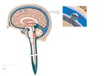
The arachnoid mater is a delicate membrane that covers the brain and forms the subarachnoid space, which contains a meshwork of collagen and elastin. Arachnoid granulations allow cerebrospinal fluid (CSF) to enter venous circulation.
Subarachnoid Space: Contains CSF and links to pia mater via trabeculae.
Arachnoid Granulations: Facilitate CSF reabsorption into blood.
Pia Mater
The pia mater is tightly attached to the brain's contours and sulci, anchored by astrocytes. It is highly vascular and forms the floor for blood vessels.
Ventricles and Cerebrospinal Fluid (CSF)
Ventricles of the Brain
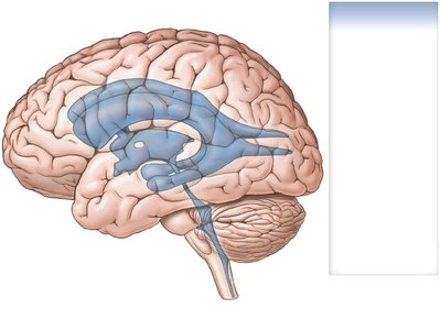
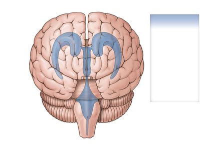
The brain contains four ventricles, which are cavities filled with CSF and lined by ependymal cells:
Lateral Ventricles: One in each cerebral hemisphere.
Third Ventricle: Located within the diencephalon.
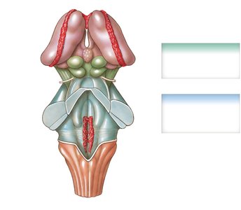
Fourth Ventricle: Between pons and cerebellum; extends into medulla oblongata.
Cerebrospinal Fluid (CSF)
CSF fills the subarachnoid space and dural sinuses, providing shock absorption, nutrient transport, and waste removal. The brain floats in CSF, preventing contact with bones.
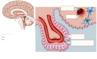
Production: CSF is produced by the choroid plexus in each ventricle.
Function: Transports nutrients, waste, and chemical messengers.
Choroid Plexus
The choroid plexus consists of specialized ependymal cells and highly permeable capillaries. Ependymal cells use active and passive transport to secrete CSF into the ventricles.
CSF Circulation
CSF is produced in the ventricles, enters the subarachnoid space via apertures, circulates throughout the brain and spinal cord, and reenters the bloodstream via arachnoid granulations.
Blood-Brain Barrier (BBB)
Structure and Function
The BBB maintains a constant environment for neurons by protecting the brain from fluctuations in blood chemistry. It is formed by special endothelial cells interconnected by tight junctions, preventing diffusion between adjacent cells. Astrocytes play a critical role in BBB function.
Selective Transport: Compounds must pass via passive or active transport.
Protection: Prevents entry of harmful substances while allowing essential nutrients.
Major Divisions: Medulla Oblongata
Structure and Function
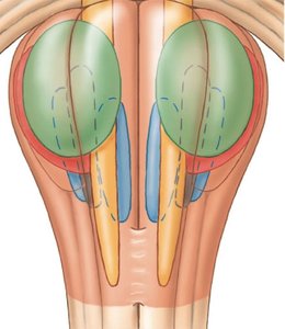
The medulla oblongata connects the spinal cord to the brain and contains nuclei that relay sensory information and regulate autonomic functions such as heart rate, blood pressure, and digestion. It also houses sensory and motor nuclei for five cranial nerves (VIII, IX, X, XI, XII).
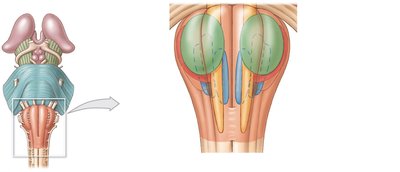
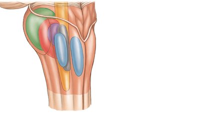
Table: Medulla Oblongata Nuclei and Functions
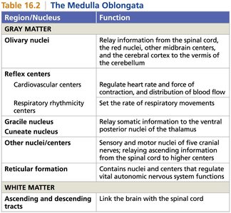
The following table summarizes the main nuclei and their functions in the medulla oblongata:
Region/Nucleus | Function |
|---|---|
Olivary nuclei | Relay information from the spinal cord, red nuclei, other midbrain centers, and cerebral cortex to the cerebellum |
Cardiovascular centers | Regulate heart rate, force of contraction, and distribution of blood flow |
Respiratory rhythmicity centers | Set the rate of respiratory movements |
Gracile nucleus | Relay somatic information to the thalamus |
Cuneate nucleus | Relay somatic information to the thalamus |
Other nuclei/centers | Sensory and motor nuclei of five cranial nerves; relay information from the spinal cord to higher centers |
Reticular formation | Contains nuclei and centers that regulate vital autonomic nervous system functions |
Ascending and descending tracts | Link the brain with the spinal cord |
Summary
Recognize the regions of the brain by their anatomical features and functions.
Understand the protective structures (meninges, CSF, BBB) and their roles.
Know the specific nuclei and their functions, especially in the medulla oblongata.
Pay attention to clinical relevance, such as injury points and autonomic regulation.
