 뒤로
뒤로Study Guide: The Cardiovascular System – Blood Vessels
스터디 가이드 - 스마트 노트
 자료에 맞춘 맞춤형 노트, 핵심 정의, 예시, 맥락을 확장해 제공합니다.
The Cardiovascular System: Blood Vessels
Overview of Blood Vessels
The cardiovascular system consists of a network of blood vessels that transport blood throughout the body. Blood vessels are classified into arteries, capillaries, and veins, each serving distinct functions in circulation.
Arteries: Carry blood away from the heart; most are oxygenated except for pulmonary arteries and fetal umbilical vessels.
Capillaries: Microscopic vessels that facilitate exchange of gases, nutrients, and wastes with tissues.
Veins: Return blood to the heart; most are deoxygenated except for pulmonary veins.
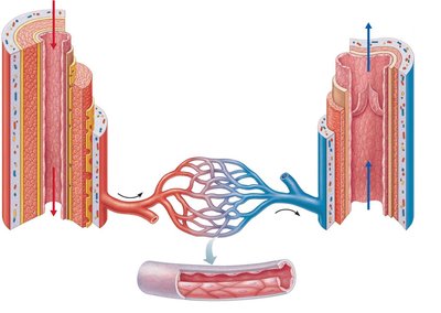
Structure of Blood Vessel Walls
Blood vessel walls are composed of three layers (tunics) in arteries and veins, while capillaries have a single layer.
Tunica intima: Innermost layer; consists of endothelium and, in larger vessels, a subendothelial connective tissue basement membrane.
Tunica media: Middle layer; composed of smooth muscle and elastin, regulated by sympathetic vasomotor nerves for vasoconstriction and vasodilation.
Tunica externa (adventitia): Outermost layer; made of collagen fibers, provides protection and reinforcement. Large vessels contain vasa vasorum to nourish the external layer.
Lumen: Central blood-containing space.
Capillaries: Consist only of endothelium with a sparse basal lamina.
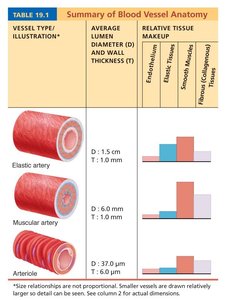
Summary of Blood Vessel Anatomy
Blood vessels differ in diameter, wall thickness, and tissue composition. The following tables summarize these differences:
Vessel Type | Average Lumen Diameter (D) | Wall Thickness (T) | Relative Tissue Makeup |
|---|---|---|---|
Elastic artery | 1.5 cm | 1.0 mm | High elastic tissue, moderate smooth muscle, moderate collagen |
Muscular artery | 6.0 mm | 1.0 mm | High smooth muscle, moderate elastic tissue, moderate collagen |
Arteriole | 37.0 μm | 1.0 μm | Moderate smooth muscle, low elastic tissue, low collagen |
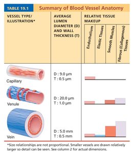
Vessel Type | Average Lumen Diameter (D) | Wall Thickness (T) | Relative Tissue Makeup |
|---|---|---|---|
Capillary | 9.0 μm | 0.5 μm | Endothelium only |
Venule | 20.0 μm | 1.0 μm | Low smooth muscle, low collagen |
Vein | 5.0 mm | 1.5 mm | Moderate smooth muscle, high collagen, low elastic tissue |
Types of Arteries
Elastic (Conducting) Arteries
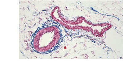
These are large, thick-walled arteries with abundant elastin in all three tunics. The aorta and its major branches are examples. Their large lumen offers low resistance, and they act as pressure reservoirs, expanding and recoiling as blood is ejected from the heart.
Muscular (Distributing) Arteries
Located distal to elastic arteries, muscular arteries deliver blood to body organs. They have a thick tunica media with more smooth muscle and are active in vasoconstriction.
Arterioles
Arterioles are the smallest arteries, leading to capillary beds. They control blood flow into capillary beds via vasodilation and vasoconstriction.
Capillaries: Structure and Function
Capillaries are microscopic vessels with walls consisting of a single layer of endothelial cells. Their small diameter allows only a single red blood cell to pass at a time. Capillaries are present in all tissues except cartilage, epithelia, cornea, and lens of the eye. Their primary function is the exchange of gases, nutrients, wastes, and hormones.
Types of Capillaries
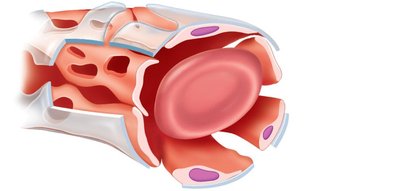
Continuous Capillaries: Most common; found in skin and muscles. Endothelial cells are connected by tight junctions, with intercellular clefts allowing passage of fluids and small solutes. In the brain, tight junctions are complete, forming the blood-brain barrier.
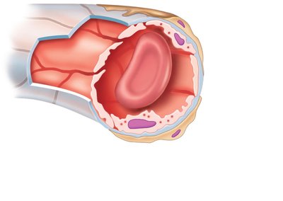
Fenestrated Capillaries: Endothelial cells contain pores (fenestrations), making them more permeable. Found in areas of absorption or filtrate formation, such as the small intestine, endocrine glands, and kidneys.
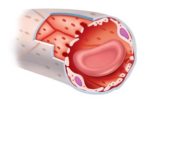
Sinusoidal Capillaries (Sinusoids): Have fewer tight junctions, larger intercellular clefts, and large lumens. Usually fenestrated, allowing passage of large molecules and blood cells. Found in the liver, bone marrow, and spleen.
Capillary Beds and Microcirculation
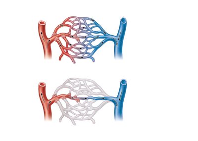
Capillary beds are interwoven networks of capillaries forming microcirculation between arterioles and venules. They consist of:
Vascular shunt: Directly connects the terminal arteriole and a postcapillary venule.
True capillaries: 10 to 100 exchange vessels per capillary bed, branching off the metarteriole or terminal arteriole.
Blood Flow Regulation
Precapillary sphincters regulate blood flow into true capillaries, controlled by local chemical conditions and vasomotor nerves.
Venous System: Venules and Veins
Venules
Venules are formed when capillary beds unite. They are very porous, allowing fluids and white blood cells to enter tissues. Postcapillary venules consist of endothelium and a few pericytes, while larger venules have one or two layers of smooth muscle cells.
Veins
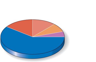
Veins are formed when venules converge. They have thinner walls and larger lumens compared to arteries. Veins are called capacitance vessels (blood reservoirs) and contain up to 65% of the blood supply. Blood pressure in veins is lower than in arteries.
Distribution of Blood
The majority of blood is found in systemic veins and venules, with smaller proportions in arteries, capillaries, and the heart.
Venous Adaptations
Large-diameter lumens offer little resistance.
Valves prevent backflow of blood, most abundant in veins of the limbs.
Venous sinuses are flattened veins with extremely thin walls (e.g., coronary sinus, dural sinuses).
Vascular Anastomoses
Vascular anastomoses are interconnections of blood vessels. Arterial anastomoses provide alternate pathways (collateral channels) to a given body region. Venous anastomoses are common, and vascular shunts of capillaries are examples of arteriovenous anastomoses.
Physiology of Circulation: Key Terms
Blood flow: Volume of blood flowing through a vessel, organ, or the entire circulation in a given period (ml/min). Equivalent to cardiac output (CO) for the entire vascular system.
Blood pressure (BP): Force per unit area exerted on the wall of a blood vessel by the blood (mm Hg). Measured as systemic arterial BP in large arteries near the heart.
Resistance (peripheral resistance): Opposition to flow; measure of the amount of friction blood encounters. Major sources: blood viscosity, vessel length, vessel diameter.
Factors Affecting Resistance
Blood viscosity: The "stickiness" of blood due to formed elements and plasma proteins.
Blood vessel length: Longer vessels increase resistance.
Blood vessel diameter: Resistance varies inversely with the fourth power of vessel radius. Small-diameter arterioles are major determinants of peripheral resistance.
Relationship Between Blood Flow, Pressure, and Resistance
Blood flow (F) is directly proportional to the pressure gradient (ΔP) and inversely proportional to resistance (R):
Formula:
Resistance is more important in influencing local blood flow because it is easily changed by altering vessel diameter.
Systemic Blood Pressure
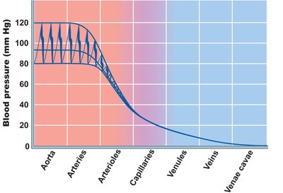
The pumping action of the heart generates blood flow. Pressure is highest in the aorta and declines throughout the pathway, reaching 0 mm Hg in the right atrium. The steepest drop occurs in arterioles.
Arterial Blood Pressure
Systolic pressure: Pressure during ventricular contraction.
Diastolic pressure: Lowest level of arterial pressure.
Pulse pressure: Difference between systolic and diastolic pressure.
Mean arterial pressure (MAP): Pressure that propels blood to tissues. Formula:
Capillary and Venous Blood Pressure
Capillary blood pressure: Ranges from 15 to 35 mm Hg; low pressure prevents rupture and facilitates exchange.
Venous blood pressure: Small pressure gradient (~15 mm Hg); low pressure due to cumulative effects of peripheral resistance.
Factors Aiding Venous Return
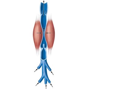
Respiratory pump: Pressure changes during breathing move blood toward the heart.
Muscular pump: Skeletal muscle contractions "milk" blood toward the heart; valves prevent backflow.
Vasoconstriction: Sympathetic control causes venoconstriction.
Maintaining Blood Pressure
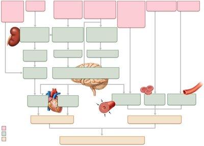
Blood pressure is regulated by the cooperation of the heart, blood vessels, kidneys, and supervision by the brain. The main factors influencing blood pressure are cardiac output (CO), peripheral resistance (PR), and blood volume.
Formula:
Blood pressure varies directly with CO, PR, and blood volume.
Cardiac Output (CO)
Determined by venous return and neural/hormonal controls.
Resting heart rate is maintained by the cardioinhibitory center via parasympathetic vagus nerves.
Stroke volume is controlled by venous return (EDV).
During stress, the cardioacceleratory center increases heart rate and stroke volume via sympathetic stimulation.
Control of Blood Pressure
Short-term neural and hormonal controls: Counteract fluctuations in blood pressure by altering peripheral resistance.
Long-term renal regulation: Counteracts fluctuations in blood pressure by altering blood volume.
Short-Term Mechanisms: Neural Controls
Neural controls maintain MAP by altering vessel diameter and blood distribution.
Operate via reflex arcs involving baroreceptors, vasomotor centers, and vascular smooth muscle.
The Vasomotor Center
Cluster of sympathetic neurons in the medulla overseeing changes in vessel diameter.
Maintains vasomotor tone (moderate constriction of arterioles).
Receives inputs from baroreceptors, chemoreceptors, and higher brain centers.
Baroreceptor-Initiated Reflexes
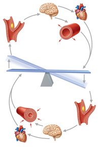
Baroreceptors are located in carotid sinuses, aortic arch, and walls of large arteries.
Increased BP stimulates baroreceptors to inhibit vasomotor center, causing vasodilation and venodilation.
Baroreceptors protect blood supply to the brain and maintain systemic BP.
Chemoreceptor-Initiated Reflexes
Chemoreceptors are located in carotid sinus, aortic arch, and large arteries of the neck.
Respond to rise in CO2, drop in pH or O2.
Increase BP via vasomotor and cardioacceleratory centers; more important in respiratory rate regulation.
Influence of Higher Brain Centers
Reflexes regulating BP are integrated in the medulla.
Higher brain centers (cortex, hypothalamus) can modify BP via relays to medullary centers.
Short-Term Mechanisms: Hormonal Controls
Adrenal medulla hormones (norepinephrine, epinephrine) cause vasoconstriction and increase cardiac output.
Angiotensin II (from kidney release of renin) causes vasoconstriction.
Atrial natriuretic peptide (ANP) causes blood volume and BP to decline, and vasodilation.
Antidiuretic hormone (ADH) causes intense vasoconstriction in cases of extremely low BP.
Long-Term Mechanisms: Renal Regulation
Baroreceptors quickly adapt to chronic high or low BP.
Long-term mechanisms control BP by altering blood volume.
Kidneys act directly (eliminate or conserve water) and indirectly (renin-angiotensin mechanism).
Direct Renal Mechanism
Increased BP or blood volume causes kidneys to eliminate more urine, reducing BP.
Decreased BP or blood volume causes kidneys to conserve water, increasing BP.
Indirect Renal Mechanism (Renin-Angiotensin)
Low arterial BP triggers release of renin.
Renin leads to production of angiotensin II, a potent vasoconstrictor.
Angiotensin II stimulates aldosterone secretion (renal reabsorption of Na+ and decreased urine formation) and ADH release.
Summary Table: Blood Vessel Types and Functions
Vessel Type | Main Function | Wall Structure |
|---|---|---|
Elastic artery | Pressure reservoir; conducts blood | Thick tunica media with abundant elastin |
Muscular artery | Distributes blood to organs | Thick tunica media with more smooth muscle |
Arteriole | Regulates blood flow to capillaries | Thin wall, mostly smooth muscle |
Capillary | Exchange of substances | Single layer of endothelium |
Venule | Collects blood from capillaries | Thin wall, some smooth muscle |
Vein | Returns blood to heart; blood reservoir | Thin tunica media, thick tunica externa |
Additional info: Academic context was added to clarify vessel functions, regulatory mechanisms, and formulas for blood flow and pressure. Tables were inferred and expanded for completeness.
