 뒤로
Study Guide: The Muscular System (Anatomy & Physiology) Chapter 10A
스터디 가이드 - 스마트 노트
 자료에 맞춘 맞춤형 노트, 핵심 정의, 예시, 맥락을 확장해 제공합니다.
The Muscular System
Muscle Actions and Interactions
The muscular system is composed of contractile tissues, including skeletal, cardiac, and smooth muscle. This section focuses on skeletal muscle, examining how muscles interact to produce movement, the criteria for naming muscles, and principles of leverage.
Muscles can only pull; they never push. Movement is achieved by the coordinated action of muscle groups.
Three main functional groups:
Prime mover (agonist): Major responsibility for producing a specific movement.
Antagonist: Opposes or reverses a particular movement. Located on the opposite side of the joint from the prime mover.
Synergist: Assists the prime mover by adding extra force or reducing unnecessary movement.
Fixator: A type of synergist that stabilizes the origin of the prime mover.
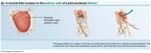
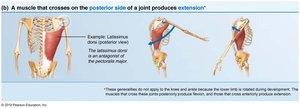
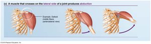
The action of a muscle can be inferred by its position relative to the joint it crosses.
Examples of Muscle Actions Relative to Joint Position
Anterior side: Muscles crossing on the anterior side of a joint produce flexion.
Posterior side: Muscles crossing on the posterior side produce extension.
Lateral side: Muscles crossing on the lateral side produce abduction.
Naming Skeletal Muscles
Skeletal muscles are named according to several criteria, which often provide clues to their location, shape, size, and function.
Location: Named for the bone or region they are associated with (e.g., temporalis over the temporal bone).
Shape: Distinctive shapes (e.g., deltoid = triangle).
Size: Terms like maximus (largest), minimus (smallest), longus (long).
Direction of fibers: Rectus (straight), transversus (right angles), oblique (angles).
Number of origins: Biceps (two origins), triceps (three origins).
Location of attachments: Named for origin and insertion (origin first).
Action: Named for the action they produce (e.g., flexor, extensor).
Multiple criteria can be combined (e.g., extensor carpi radialis longus).
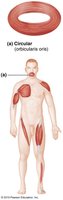
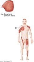
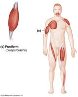
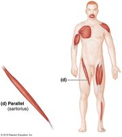
Fascicle Arrangements in Skeletal Muscles
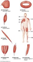
All skeletal muscles consist of fascicles (bundles of muscle fibers). The arrangement of fascicles determines the muscle's shape and functional capabilities.
Circular: Fascicles arranged in concentric rings (e.g., orbicularis oris).
Convergent: Broad origin, fascicles converge toward a single tendon (e.g., pectoralis major).
Parallel: Fascicles parallel to the long axis of the muscle (e.g., sartorius).
Fusiform: Spindle-shaped with parallel fibers (e.g., biceps brachii).
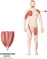
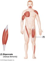
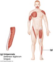
Pennate: Short fascicles attach obliquely to a central tendon.
Unipennate: Fascicles attach to one side (e.g., extensor digitorum longus).
Bipennate: Fascicles attach to both sides (e.g., rectus femoris).
Multipennate: Multiple fascicles attach to one tendon (e.g., deltoid).
Major Skeletal Muscles of the Body
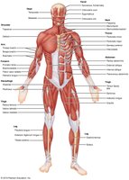
There are over 600 skeletal muscles in the human body, grouped by function and location. Muscle tables typically include description, origin and insertion, actions, and innervation.
Description: Location relative to other muscles.
Origin and insertion: Usually a joint between these points.
Actions: Movement caused by contraction.
Innervation: Major nerve supplying the muscle.
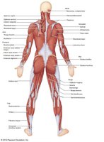
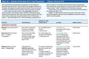
Muscles of the Head: Facial Expression
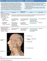
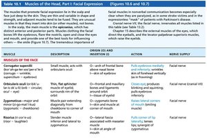
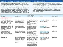
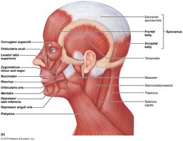
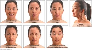
Muscles of facial expression are unique because they insert into the skin, not bone, and are important for nonverbal communication. All are innervated by cranial nerve VII (facial nerve).
Two groups:
Muscles of the scalp
Muscles of the face
Muscle | Description | Origin & Insertion | Action | Nerve Supply |
|---|---|---|---|---|
Corrugator supercilii | Small muscle; activity causes eyebrows to move medially and downward | Arch of frontal bone above nasal bone; skin of eyebrow | Pulls eyebrows medially and downward | Facial nerve |
Orbicularis oculi | Thin, flat sphincter muscle of eyelid | Frontal and maxillary bones; skin around eyelids | Closes eyes | Facial nerve |
Zygomaticus (major and minor) | Muscle pair extending diagonally from cheekbone to corner of mouth | Zygomatic bone; skin and muscle at corner of mouth | Raises lateral corners of mouth (smiling) | Facial nerve |
Risorius | Slender muscle; runs inferior and lateral to zygomaticus | Fascia of masseter muscle; skin at angle of mouth | Pulls corner of lip laterally | Facial nerve |
Muscles of Mastication and Tongue Movement
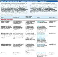
Four pairs of muscles are involved in mastication (chewing), all innervated by cranial nerve V. The tongue is anchored and moved by three extrinsic muscles.
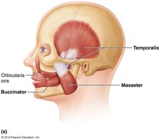
Prime movers of jaw closure: Temporalis and masseter
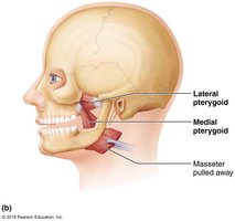
Grinding movements: Pterygoids
Chewing role: Buccinator
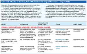
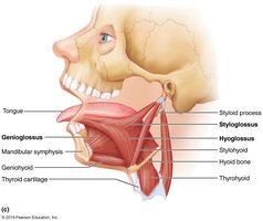
Tongue movement: Genioglossus, hyoglossus, styloglossus
Muscle | Description | Origin & Insertion | Action | Nerve Supply |
|---|---|---|---|---|
Temporalis | Fan-shaped muscle covering parts of temporal, frontal, and parietal bones | Temporal fossa; coronoid process of mandible | Closes jaw | Trigeminal nerve |
Masseter | Powerful muscle covering lateral aspect of mandibular ramus | Zygomatic arch; angle and ramus of mandible | Closes jaw | Trigeminal nerve |
Medial pterygoid | Deep two-headed muscle; runs along internal surface of mandible | Medial surface of lateral pterygoid plate; medial surface of mandible | Grinding movements | Trigeminal nerve |
Lateral pterygoid | Deep two-headed muscle; runs along internal surface of mandible | Lateral surface of lateral pterygoid plate; condylar process of mandible | Grinding movements | Trigeminal nerve |
Buccinator | Horizontal muscle; forms cheek | Mandible and maxilla; orbicularis oris | Compresses cheek | Facial nerve |
Muscles of the Anterior Neck and Throat: Swallowing
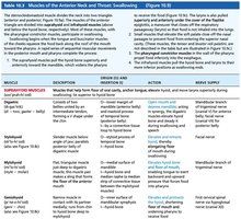
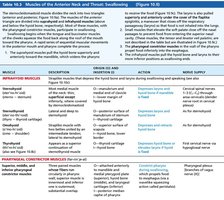
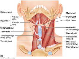
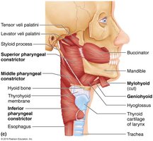
The sternocleidomastoid divides the neck into anterior and posterior triangles. Muscles are grouped as suprahyoid and infrahyoid based on their relation to the hyoid bone. These muscles facilitate swallowing by moving the hyoid bone and larynx.
Suprahyoid muscles: Four deep muscles that elevate the hyoid bone and move the larynx during swallowing.
Infrahyoid muscles: Four straplike muscles that depress the hyoid bone and larynx during swallowing and speaking.
Muscle | Description | Origin & Insertion | Action | Nerve Supply |
|---|---|---|---|---|
Mylohyoid | Flat, triangular muscle forms floor of oral cavity | Medial surface of mandible; hyoid bone | Elevates hyoid bone and floor of mouth | Mandibular branch of trigeminal nerve |
Digastric | Two bellies united by intermediate tendon | Lower margin of mandible; hyoid bone | Opens mouth and depresses mandible | Mandibular branch of trigeminal nerve; facial nerve |
Sternohyoid | Most medial muscle of neck | Manubrium and medial end of clavicle; hyoid bone | Depresses hyoid bone | Ansa cervicalis (C1-C3) |
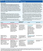
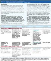
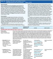
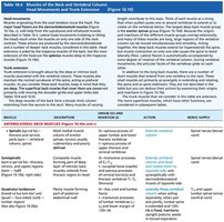
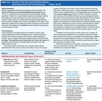
Muscles of the Neck and Vertebral Column: Head Movements and Trunk Extension
Muscles in this region are divided into anterolateral neck muscles (move the head) and intrinsic muscles of the back (extend the trunk and maintain posture).
Anterolateral neck muscles: Move the head.
Intrinsic back muscles: Extend the trunk and maintain posture.
Muscle | Description | Origin & Insertion | Action | Nerve Supply |
|---|---|---|---|---|
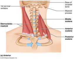
Sternocleidomastoid | Two-headed muscle; key landmark in neck | Manubrium of sternum and medial portion of clavicle; mastoid process of temporal bone | Flexes and laterally rotates the head | Accessory nerve (cranial nerve XI) |
Scalenes | Three muscles (anterior, middle, posterior) located laterally on neck | Transverse processes of cervical vertebrae; first two ribs | Elevate first two ribs; flex and rotate neck | Cervical spinal nerves |
Splenius | Broad, two-part muscle; extends or hyperextends head | Ligamentum nuchae and spinous processes of vertebrae; mastoid process and occipital bone | Extend or hyperextend head | Cervical spinal nerves |
Summary Table: Functional Groups of Skeletal Muscles
Group | Function | Example |
|---|---|---|
Prime mover (agonist) | Produces specific movement | Biceps brachii (elbow flexion) |
Antagonist | Opposes or reverses movement | Triceps brachii (elbow extension) |
Synergist | Assists prime mover | Brachialis (assists elbow flexion) |
Fixator | Stabilizes origin | Muscles stabilizing scapula |
Additional info: Academic context was added to expand brief points and tables, ensuring completeness and clarity for exam preparation.
