 뒤로
Study Guide: The Muscular System (Anatomy & Physiology, Chapter 11)
스터디 가이드 - 스마트 노트
 자료에 맞춘 맞춤형 노트, 핵심 정의, 예시, 맥락을 확장해 제공합니다.
The Muscular System
Introduction to the Muscular System
The muscular system consists exclusively of skeletal muscles, which are responsible for movement, posture, and heat production. The organization of muscles affects their power, range, and speed of movement.
Fascicle Arrangement
Types of Fascicle Arrangement
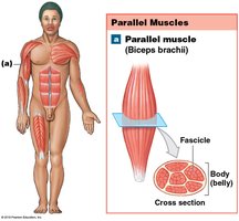
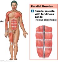
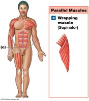
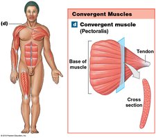
Skeletal muscle fibers are grouped into bundles called fascicles. The arrangement of these fascicles determines the muscle's function and efficiency. Muscles are classified based on fascicle arrangement:
Parallel muscles: Fascicles run parallel to the long axis of the muscle. These muscles can be flat or cylindrical with a central belly. Example: biceps brachii. Tension depends on the number of myofibrils.
Convergent muscles: Muscle fibers spread out like a fan and converge on a single attachment site (tendon, aponeurosis, or raphe). Example: pectoralis major. Fibers can pull in different directions.
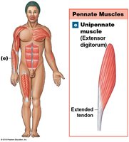
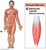
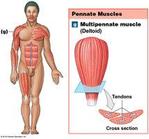
Pennate muscles: Fascicles are arranged at an angle to the tendon. These muscles develop more tension but do not move their tendons as far. Types include unipennate (e.g., extensor digitorum), bipennate (e.g., rectus femoris), and multipennate (e.g., deltoid).
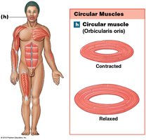
Circular muscles (sphincters): Fascicles are arranged in concentric rings, acting as valves to control openings. Example: orbicularis oris.
Levers in the Muscular System
Classes of Levers
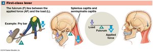
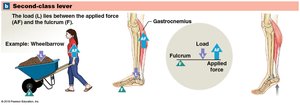
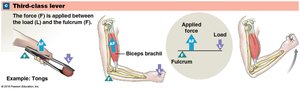
Muscles attach to bones, which act as levers. The site of attachment affects force, speed, and range of movement. Each joint acts as a fulcrum. Levers can change the direction, distance, speed, and strength of applied force. There are three classes of levers:
First-class lever: The fulcrum is between the applied force and the load. Example: extension of the neck and lifting the head.
Second-class lever: The load is between the applied force and the fulcrum. Example: ankle extension (plantar flexion) by calf muscles.
Third-class lever: The applied force is between the load and the fulcrum. This is the most common lever in the body, maximizing speed and distance at the expense of force. Example: flexion at the elbow.
Origins, Insertions, and Actions of Muscles
Muscle Attachments and Actions
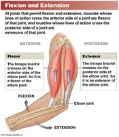
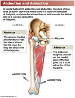
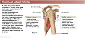
Muscles attach to bones at two points: the origin (fixed, usually proximal) and the insertion (movable, usually distal). The action of a muscle is the movement produced by its contraction, described in terms of its effect on bones or joints (e.g., flexion, extension, adduction).
Agonist (prime mover): Responsible for a specific movement.
Antagonist: Opposes the action of the agonist.
Synergist: Assists the agonist.
Fixator: Stabilizes the origin of the agonist.
Agonists and antagonists work in pairs; when one contracts, the other stretches (e.g., flexors and extensors).
Naming Skeletal Muscles
Muscle Naming Conventions
Muscle names provide descriptive information about their location, appearance, or function. Common naming criteria include:
Region of the body: e.g., temporalis (temple), brachial (arm), femoris (thigh).
Position, direction, or fascicle arrangement: e.g., externus (superficial), internus (deep), transversus (across), oblique (slanting), rectus (straight).
Structural characteristics: Origin and insertion (e.g., genioglossus), number of tendons (e.g., biceps, triceps), shape and size (e.g., deltoid, trapezius).
Action: e.g., flexor, extensor, abductor, adductor, pronator, supinator.
Some names reflect occupations or habits (e.g., buccinator means "trumpeter").
Axial and Appendicular Muscles
Muscle System Divisions
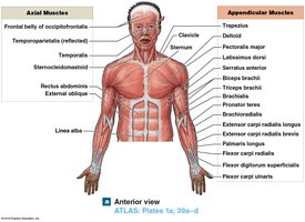
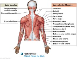
The muscular system is divided into axial and appendicular muscles:
Axial muscles: Constitute 60% of skeletal muscles. They position the head and vertebral column, move the rib cage, and form the pelvic floor.
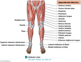
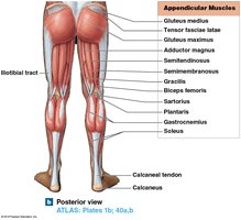
Appendicular muscles: Move and support the pectoral and pelvic girdles and limbs.
Principal Axial Muscles
Muscles of the Head and Neck
Axial muscles are grouped by location and function:
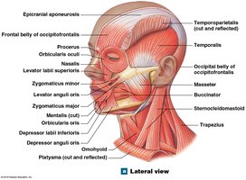
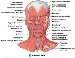
Muscles of facial expression: Originate on the skull. Examples: orbicularis oris (mouth), buccinator (cheek).
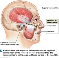
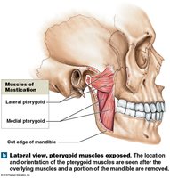
Muscles of mastication: Move the mandible. Examples: masseter, temporalis, pterygoid muscles.
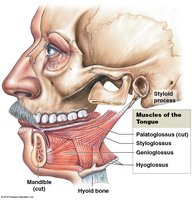
Muscles of the tongue: Names end in "glossus". Examples: palatoglossus, styloglossus, genioglossus, hyoglossus.
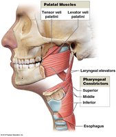
Muscles of the pharynx: Initiate swallowing. Examples: pharyngeal constrictors, palatal muscles, laryngeal elevators.
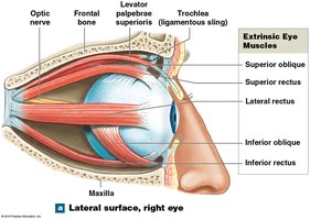
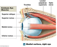
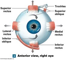
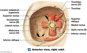
Extrinsic eye muscles: Control eye position. Examples: inferior rectus, medial rectus, superior rectus, lateral rectus, inferior oblique, superior oblique.
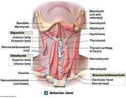
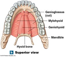
Muscles of the anterior neck: Control larynx position, depress mandible, and support tongue and pharynx. Examples: digastric, mylohyoid, geniohyoid, stylohyoid, sternocleidomastoid, omohyoid.
Muscles of the Vertebral Column
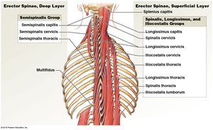
These muscles are divided into superficial and deep layers:
Erector spinae (superficial): Spinalis, longissimus, and iliocostalis groups.
Erector spinae (deep): Semispinalis, multifidus, interspinales, intertransversarii, rotatores.
Spinal flexors: Longus capitis, longus colli (neck); quadratus lumborum (lumbar region).
Oblique and Rectus Muscles
Oblique muscles compress underlying structures and rotate the vertebral column. Rectus muscles flex the vertebral column and oppose erector spinae. The diaphragm is a major muscle used in breathing, dividing the thoracic and abdominopelvic cavities.
Muscles of the Pelvic Floor
These muscles support pelvic organs, flex the sacrum and coccyx, and control movement through the urethra and anus. The perineum is divided into the urogenital and anal triangles, with the pelvic diaphragm forming the foundation of the anal triangle.
Principal Appendicular Muscles
Muscles of the Shoulders and Upper Limbs
Appendicular muscles position and stabilize the pectoral and pelvic girdles and move the limbs. Major groups include:
Muscles that position the pectoral girdle: Trapezius, serratus anterior, subclavius, pectoralis minor, rhomboid major/minor, levator scapulae.
Muscles that move the arm: Deltoid, supraspinatus, subscapularis, teres major/minor, infraspinatus, coracobrachialis, pectoralis major, latissimus dorsi.
Rotator cuff: Supraspinatus, infraspinatus, teres minor, subscapularis.
Muscles that move the forearm and hand: Biceps brachii, triceps brachii, brachialis, brachioradialis, supinator, pronator teres, pronator quadratus.
Muscles that move the fingers: Extrinsic (forearm) and intrinsic (hand) muscles.
Muscles of the Pelvis and Lower Limbs
The pelvic girdle is tightly bound to the axial skeleton, permitting little movement. Major groups include:
Muscles that move the thigh: Gluteal muscles (gluteus maximus, medius, minimus), tensor fasciae latae, lateral rotators (piriformis, obturator), adductors (pectineus, adductor brevis/longus/magnus, gracilis), iliopsoas (psoas major, iliacus).
Muscles that move the leg: Flexors of the knee (hamstrings: biceps femoris, semitendinosus, semimembranosus, sartorius, popliteus), knee extensors (quadriceps femoris: vastus muscles, rectus femoris).
Muscles that move the foot and toes: Extrinsic muscles (gastrocnemius, soleus, fibularis, tibialis posterior, tibialis anterior, extensor digitorum longus, extensor hallucis longus, flexor digitorum longus, flexor hallucis longus), intrinsic muscles (originate on tarsal and metatarsal bones).
Effects of Exercise and Integration with Other Body Systems
Functional Relationships and Exercise
The muscular system is supported by other body systems:
Cardiovascular system: Delivers oxygen and nutrients, removes carbon dioxide.
Respiratory system: Responds to oxygen demand of muscles.
Integumentary system: Disperses heat from muscle activity.
Nervous and endocrine systems: Direct responses of all systems.
Exercise produces various responses in these systems, enhancing overall health and function.
Additional info: This study guide covers the major topics and subtopics from Chapter 11 of a college-level Anatomy & Physiology course, including muscle structure, function, naming conventions, and integration with other body systems.
