 뒤로
뒤로Study Guide: The Nervous System (Anatomy & Physiology)
스터디 가이드 - 스마트 노트
 자료에 맞춘 맞춤형 노트, 핵심 정의, 예시, 맥락을 확장해 제공합니다.
The Nervous System
Overview and Functional Divisions
The nervous system is responsible for monitoring internal and external environments, integrating sensory information, and coordinating responses between organs. It is divided into the central nervous system (CNS) and peripheral nervous system (PNS).
Central Nervous System (CNS): Composed of the brain and spinal cord; processes sensory data and issues motor commands.
Peripheral Nervous System (PNS): All nervous tissue outside the CNS; delivers sensory data and motor commands.
Afferent Division: Brings sensory data to the CNS.
Efferent Division: Carries motor commands to tissues.
Somatic Nervous System: Controls skeletal muscle.
Autonomic Nervous System: Regulates smooth/cardiac muscle, glands, and adipose tissue. Includes sympathetic (fight or flight), parasympathetic (rest and digest), and enteric (digestive functions) divisions.
Neurons: Structure and Classification
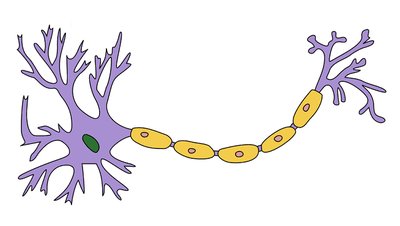
Neurons are the basic functional units of the nervous system, possessing excitable membranes. Most neurons cannot divide after injury or disease.
Cell Body (Soma): Contains the nucleus.
Dendrites: Receive information from other cells.
Axon: Carries information to other cells.
Axon Terminals: Endings where neurotransmitters are released.
Myelin Sheath: Insulates axons, increasing conduction speed.
Structural Classification of Neurons
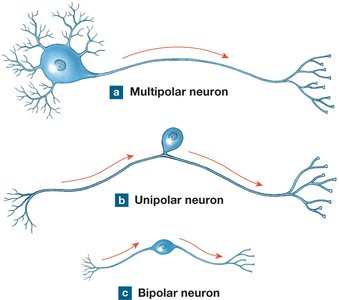
Neurons are classified based on their structure:
Multipolar: Multiple dendrites, single axon; most common.
Unipolar: Dendrites and axon are continuous.
Bipolar: One dendrite and one axon.
Functional Classification
Sensory Neurons: Form the afferent division; monitor external environment, proprioception, and internal organ systems.
Motor Neurons: Control effectors; somatic (skeletal muscle) and visceral (organs).
Interneurons: Located between sensory and motor neurons; interconnect nerve cells.
Neuroglia: Supporting Cells
Neuroglia protect neurons, provide structural framework, act as phagocytes, and regulate the internal environment.
Astrocytes: Maintain blood-brain barrier, repair tissue, guide development, control environment.
Oligodendrocytes: Form myelin sheath in CNS.
Ependymal Cells: Line cavities, produce cerebrospinal fluid (CSF).
Microglia: Phagocytic cells in CNS.
Satellite Cells: Surround neuron cell bodies in PNS.
Schwann Cells: Form myelin sheath in PNS.
Membrane Potential and Action Potentials
Resting Membrane Potential
The difference in charge across the neuronal membrane is the membrane potential (), typically -70 mV.
Ion Concentrations: Extracellular and intracellular fluids contain different ions.
Equilibrium Potential: Each ion has its own equilibrium potential.
Channels and Transporters
Voltage-Gated Ion Channels: Open/close in response to membrane potential changes.
Graded Potential: Temporary change in membrane potential; varies with stimulus size.
Action Potential: Occurs if threshold is reached; follows all-or-nothing principle.
Stages of Action Potential
Depolarization: Voltage-gated Na+ channels open, Na+ enters, becomes more positive. Equilibrium potential: +60 mV.
Repolarization: Na+ channels inactivate, K+ channels open, K+ leaves, becomes more negative. Equilibrium potential: -90 mV.
Hyperpolarization: K+ channels close, drops below resting before returning to normal.
Refractory Periods
Absolute Refractory Period: Cell cannot respond to further stimuli; Na+ channels are open/inactive.
Relative Refractory Period: Stronger stimulus required; Na+ channels are closed.
Propagation of Action Potentials
Continuous Propagation: Slow, occurs in unmyelinated axons.
Saltatory Propagation: Fast, action potential jumps between nodes of Ranvier in myelinated axons.
Speed: Fastest in large-diameter, myelinated axons; high-priority information uses these fibers.
Synapses and Neurotransmitters
Types of Synapses
Electrical Synapse: Direct physical contact; ions pass between cells.
Chemical Synapse: Neurotransmitter release; includes acetylcholine (excitatory), norepinephrine (excitatory), dopamine (inhibitory), GABA (inhibitory), serotonin (inhibitory).
Synaptic Transmission
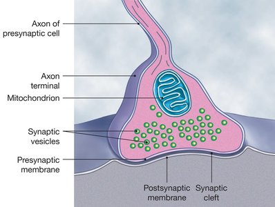
Neurotransmitters released from axon terminal.
Postsynaptic cell can be neuron, muscle, or secretory cell.
Neurotransmitters diffuse across synaptic cleft, bind to receptors, trigger graded potential.
Enzymatic breakdown and reuptake of neurotransmitter.
Central Nervous System Organization
Meninges
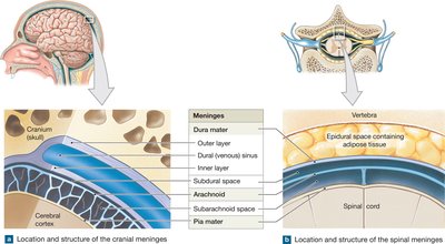
The CNS is surrounded by three layers of specialized membranes called meninges, providing stability and shock absorption.
Dura Mater: Outermost layer; fused to skull, forms dural folds and sinuses.
Arachnoid Mater: Middle layer; subarachnoid space contains CSF.
Pia Mater: Innermost layer; highly vascular, bound to brain.
Epidural Space: Between vertebra and dura mater; filled with adipose tissue.
Major Regions of the Brain
The brain consists of six major regions:
Cerebrum: Largest portion; conscious thought, memory, sensations.
Diencephalon: Relay centers; includes thalamus, hypothalamus, epithalamus.
Midbrain: Processes visual/auditory information; regulates motor output.
Pons: Connects cerebellum to brainstem; cranial nerve nuclei, respiration.
Medulla Oblongata: Connects brain to spinal cord; regulates heart rate, blood flow, respiration.
Cerebellum: Balance, fine-tuning movements.
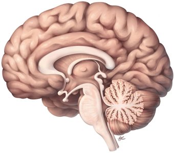
Brain Organization
Nuclei: Clusters of cell bodies (gray matter); processing centers.
Tracts: Bundles of axons (white matter); signal transmission.
Corpus Callosum: Tract connecting cerebral hemispheres.
Basal Nuclei: Islands of gray matter within the brain.
Ventricular System and Cerebrospinal Fluid (CSF)
CSF is found in brain ventricles, produced by the choroid plexus, and cushions/supports the CNS.
Ventricles: Lined by ependymal cells; fourth ventricle continuous with spinal cord central canal.
CSF Flow: Enters subarachnoid space via fourth ventricle; flows throughout subarachnoid space; excess returns to venous circulation via arachnoid granulations.
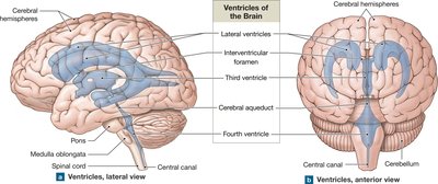
Cerebrum and Functional Areas
Primary Motor Cortex: Directs voluntary movements.
Primary Somatosensory Cortex: Receives touch, pressure, pain, temperature information.
Association Areas: Interpret sensory/motor information.
Lobes: Frontal, parietal, occipital, temporal, insula; each with specialized functions.
Diencephalon
Epithalamus: Contains choroid plexus, pineal gland (melatonin).
Thalamus: Relay center for sensory information (except olfactory).
Hypothalamus: Emotions, autonomic function, hormones, behavioral drives.
Midbrain, Pons, Medulla Oblongata, Cerebellum
Midbrain: Visual/auditory processing, motor regulation.
Pons: Connects cerebellum, cranial nerve nuclei, respiration.
Medulla Oblongata: Heart rate, blood flow, respiration regulation.
Cerebellum: Balance, movement coordination.
Spinal Cord Organization
Structure and Function
The spinal cord carries sensory and motor information between the brain and body.
Regions correspond to vertebrae: 8 cervical, 12 thoracic, 5 lumbar, 5 sacral, 1 coccygeal.
Cervical/lumbosacral enlargements provide innervation to arms/legs.
CSF found in central canal.
White matter surrounds gray matter; spinal ganglia contain sensory cell bodies.
Posterior roots: sensory axons; anterior roots: motor axons; spinal nerves: mixed.
Sensory and Motor Pathways
Sensory Pathways
Sensory pathways carry information to the CNS.
First-Order Neuron: Delivers sensations directly to CNS.
Second-Order Neuron: Interneuron receives signal from first-order neuron.
Third-Order Neuron: Relays information in the thalamus.
Posterior Column Pathway: Example of sensory pathway; involves decussation (crossing over).
Motor Pathways
Motor pathways issue commands from CNS to effectors.
Corticospinal Pathway: Voluntary control of skeletal muscles.
Upper Motor Neurons: Cell body in primary motor cortex.
Lower Motor Neurons: Cell body in brainstem or spinal cord.
Reflex Arcs
Components and Types
Reflexes are automatic responses to stimuli, typically removing or opposing the stimulus.
Stimulus activates receptor.
Sensory neuron is activated.
Information processed in CNS (interneuron).
Motor neurons activated.
Response occurs.
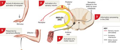
Monosynaptic Reflex: Sensory neuron synapses directly on motor neuron (e.g., stretch reflex).
Polysynaptic Reflex: Involves interneurons; more delay, greater response (e.g., flexor reflex).
Innate Reflexes: Inborn, result from developmental connections.
Acquired Reflexes: Learned motor patterns, result from repetition.
Autonomic Nervous System
Organization and Function
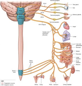
The autonomic nervous system (ANS) controls involuntary functions of visceral organs, maintaining homeostasis.
Preganglionic Neurons: Visceral neurons in brainstem/spinal cord; synapse on postganglionic neurons.
Postganglionic Neurons: Act on effectors.
Cholinergic Fibers: Release acetylcholine; preganglionic fibers always excitatory.
Adrenergic Fibers: Release norepinephrine; postganglionic sympathetic fibers typically excitatory.
Sympathetic Division
Fight or Flight: Increases metabolism, respiratory rate, heart rate, blood pressure; decreases digestive/urinary functions.
Short preganglionic fibers originate in thoracolumbar region; synapse near spinal cord; long postganglionic fibers synapse in target organ.
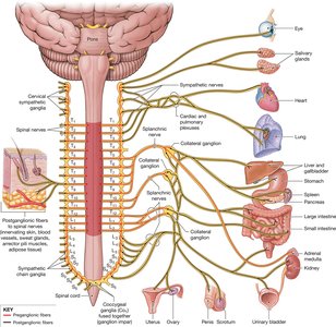
Parasympathetic Division
Rest and Digest: Decreases metabolism, heart rate, blood pressure; increases digestive/urinary functions.
Long preganglionic neurons originate in brainstem/sacral regions; synapse near target organs; short postganglionic neurons.
Summary Table: Structural and Functional Divisions of the Nervous System
Division | Structure | Function |
|---|---|---|
CNS | Brain, spinal cord | Process sensory data, issue motor commands |
PNS | Nerves outside CNS | Deliver sensory/motor information |
Somatic | Skeletal muscle | Voluntary movement |
Autonomic | Visceral organs | Involuntary regulation |
Sympathetic | Thoracolumbar | Fight or flight |
Parasympathetic | Brainstem, sacral | Rest and digest |
Key Equations
Membrane Potential:
Nernst Equation (for equilibrium potential):
Review Questions
Which cells maintain the blood-brain barrier?
Which ion is responsible for hyperpolarization?
Why do action potentials always move forward?
How does the membrane return to normal following an action potential?
Which structure of the brain produces cerebrospinal fluid?
Which lobe of the brain is involved in sight/visual processing?
What structures of the brain assist in balance and coordination?
Where are sensory cell bodies located?
Which side of the motor cortex would control the right arm?
Would sweating be a sympathetic or parasympathetic response?
In which division do we have long preganglionic fibers?
Where do parasympathetic fibers originate?
Additional info: Academic context and expanded explanations were added to ensure completeness and clarity for exam preparation.
