 뒤로
Study Guide: The Respiratory System (Anatomy & Physiology)
스터디 가이드 - 스마트 노트
 자료에 맞춘 맞춤형 노트, 핵심 정의, 예시, 맥락을 확장해 제공합니다.
The Respiratory System: Overview
Major Functions and Processes
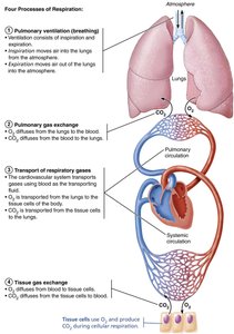
The respiratory system is essential for gas exchange, supplying oxygen (O2) to cells and removing carbon dioxide (CO2) produced during cellular respiration. It works closely with the cardiovascular system to ensure efficient transport and exchange of gases throughout the body.
Gas Exchange: The primary function is to supply O2 and remove CO2.
Processes of Respiration:
Pulmonary ventilation: Movement of air into and out of the lungs (breathing).
Pulmonary gas exchange: Exchange of O2 and CO2 between lungs and blood.
Transport of respiratory gases: Movement of O2 and CO2 in blood.
Tissue gas exchange: Exchange of O2 and CO2 between blood and tissues.
Other Functions: Olfaction (smell) and speech.
Functional Anatomy of the Respiratory System
Major Organs and Divisions
The respiratory system is divided into upper and lower regions, each with distinct structures and functions.
Upper Respiratory System: Nose, paranasal sinuses, pharynx
Lower Respiratory System: Larynx, trachea, bronchi (and branches), lungs, alveoli
Respiratory Muscles: Classified as part of the muscular system (e.g., diaphragm, intercostals)
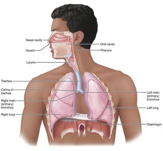
Table: Upper Respiratory System Structures
Structure | Description | Function |
|---|---|---|
Nose | External portion supported by bone and cartilage; internal cavity divided by septum, lined with mucosa | Produces mucus; filters, warms, moistens air; resonance for speech; olfactory receptors |
Paranasal sinuses | Mucosa-lined, air-filled cavities in cranial bones | Lighten skull; warm, moisten, filter air |
Pharynx | Passageway connecting nasal cavity to larynx and oral cavity to esophagus; three regions | Passageway for air and food; houses tonsils; exposes immune system to inhaled antigens |
Table: Lower Respiratory System Structures
Structure | Description | Function |
|---|---|---|
Larynx | Connects pharynx to trachea; framework of cartilage and connective tissue | Air passageway; prevents food entry; voice production |
Trachea | Flexible tube from larynx dividing into main bronchi; C-shaped cartilage rings | Air passageway; cleans, warms, moistens air |
Bronchial tree | Main bronchi subdivide into smaller branches; bronchioles lack cartilage, have smooth muscle | Air passageways; cleans, warms, moistens air |
Alveoli | Microscopic chambers at bronchial termini; simple squamous epithelium | Main sites of gas exchange; surfactant reduces surface tension |
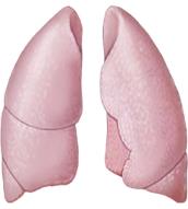
Lungs | Paired organs flanking mediastinum; composed of alveoli and passageways | House respiratory passages; elastic tissue allows recoil |
Pleurae | Serous membranes lining thoracic cavity and covering lungs | Produce lubricating fluid; compartmentalize lungs |
The Nose and Paranasal Sinuses
Structure and Function
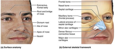
The nose is the only externally visible part of the respiratory system. It provides an airway, moistens and warms air, filters and cleans inspired air, serves as a resonating chamber for speech, and houses olfactory receptors.
External Nose: Surface features include root, bridge, dorsum nasi, apex, and nostrils (nares). Skeletal framework consists of nasal/frontal bones, maxillary bones, and hyaline cartilage.
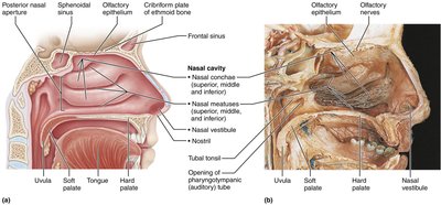
Nasal Cavity: Divided by nasal septum; lined with mucosa (olfactory and respiratory); contains nasal conchae and meatuses to increase surface area and enhance airflow.
Paranasal Sinuses: Located in frontal, sphenoid, ethmoid, and maxillary bones; lighten skull, warm/moisten air, secrete mucus.
The Pharynx
Regions and Functions
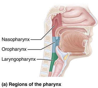
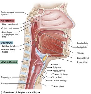
The pharynx is a funnel-shaped muscular tube connecting the nasal cavity to the larynx and the mouth to the esophagus. It is divided into three regions:
Nasopharynx: Posterior to nasal cavity; lined with ciliated epithelium; serves as airway; contains pharyngeal tonsils.
Oropharynx: Posterior to oral cavity; passageway for food and air; lined with stratified squamous epithelium; contains palatine and lingual tonsils.
Laryngopharynx: Posterior to larynx; passageway for food and air; lined with stratified squamous epithelium; continuous with esophagus.
The Larynx
Structure and Function
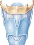
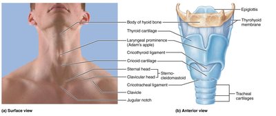
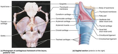
The larynx (voice box) extends from the pharynx to the trachea and is composed of nine cartilages. It provides an open airway, routes air and food, and houses vocal folds for sound production.
Cartilages: Includes thyroid (Adam's apple), cricoid, epiglottis (elastic cartilage), arytenoid, cuneiform, and corniculate.
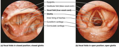
Vocal Folds: True vocal cords vibrate to produce sound; vestibular folds (false vocal cords) help close glottis during swallowing.
Epithelium: Stratified squamous above vocal folds; ciliated columnar below.
The Trachea
Structure and Histology
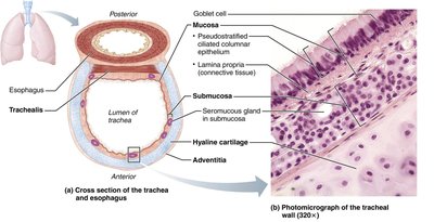
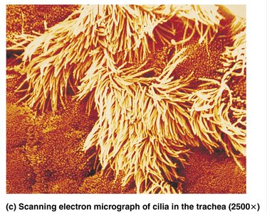
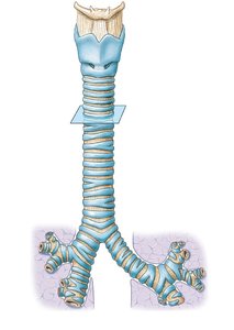
The trachea (windpipe) is a flexible tube supported by C-shaped cartilage rings. It conducts air from the larynx to the bronchi and is lined with ciliated pseudostratified epithelium containing goblet cells.
Layers: Mucosa (epithelium), submucosa (glands, cartilage), adventitia (outer connective tissue).
Trachealis Muscle: Smooth muscle that contracts during coughing.
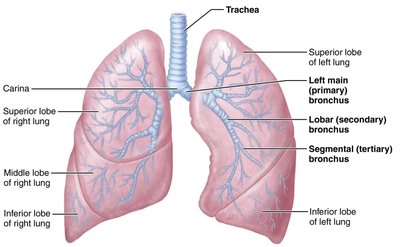
Carina: Last tracheal cartilage marking the division into main bronchi.
The Bronchi and Subdivisions
Bronchial Tree and Respiratory Zones
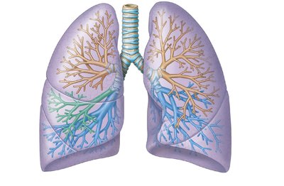
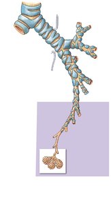
The bronchial tree consists of branching airways from the trachea to the alveoli. Structural changes occur as bronchi branch into bronchioles, including loss of cartilage and increased smooth muscle.
Conducting Zone: Trachea → main bronchi → lobar bronchi → segmental bronchi → bronchioles → terminal bronchioles.
Respiratory Zone: Terminal bronchioles → respiratory bronchioles → alveolar ducts → alveolar sacs (sites of gas exchange).
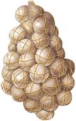
Alveoli: Clusters of thin-walled sacs surrounded by capillaries; type I cells (gas exchange), type II cells (surfactant production), macrophages (defense).
Summary Table: Bronchial Tree Branching
Structure | Diameter | Cartilage | Epithelium | Function |
|---|---|---|---|---|
Main Bronchi | ~1 cm | Rings | Pseudostratified | Air passage |
Lobar Bronchi | ~5 mm | Plates | Columnar | Air passage |
Segmental Bronchi | ~1 mm | None | Cuboidal | Air passage |
Bronchioles | <1 mm | None | Cuboidal | Air passage |
Alveoli | Microscopic | None | Squamous | Gas exchange |
Clinical Correlations
Common Disorders
Rhinitis: Inflammation of nasal mucosa; can spread to sinuses and cause headaches.
Laryngitis: Inflammation of vocal folds; causes hoarseness.
Tracheal Obstruction: Life-threatening; Heimlich maneuver can expel blockage.
Asthma: Reversible airway obstruction; treated with anti-inflammatory drugs.
COPD: Chronic obstructive pulmonary disease; includes emphysema and chronic bronchitis.
Key Terms and Concepts
Pulmonary Ventilation: Movement of air in and out of lungs.
Gas Exchange: Diffusion of O2 and CO2 across membranes.
Surfactant: Substance produced by type II alveolar cells to reduce surface tension.
Respiratory Membrane: Thin barrier for gas exchange between alveoli and capillaries.
Dead Space: Volume of air not involved in gas exchange.
Example: Boyle's Law in Pulmonary Ventilation
Boyle's Law describes the relationship between pressure and volume in the lungs:
As lung volume increases during inspiration, pressure decreases, allowing air to flow in. During expiration, volume decreases and pressure increases, forcing air out.
Summary Table: Pulmonary Volumes and Capacities
Volume/Capacity | Definition | Average Value (Adult Male) |
|---|---|---|
Tidal Volume (TV) | Air moved per breath | 500 mL |
Inspiratory Reserve Volume (IRV) | Extra air inhaled | 3300 mL |
Expiratory Reserve Volume (ERV) | Extra air exhaled | 1000 mL |
Residual Volume (RV) | Air remaining after exhalation | 1200 mL |
Vital Capacity (VC) | TV + IRV + ERV | 4800 mL |
Total Lung Capacity (TLC) | Sum of all volumes | 6000 mL |
Additional info:
Surfactant is critical for preventing alveolar collapse, especially in premature infants.
Respiratory diseases such as tuberculosis and lung cancer are major causes of morbidity and mortality.
Sleep apnea is a common disorder affecting breathing during sleep, often treated with CPAP devices.
