 뒤로
Study Guide: The Skeletal System (Anatomy & Physiology)
스터디 가이드 - 스마트 노트
 자료에 맞춘 맞춤형 노트, 핵심 정의, 예시, 맥락을 확장해 제공합니다.
The Skeletal System
Overview and Components
The skeletal system is a fundamental organ system in human anatomy, providing structure, protection, and facilitating movement. It consists of bones, joints, cartilages, and ligaments, and is divided into two main subdivisions: the axial skeleton and the appendicular skeleton.
Bones: Provide structural support and serve as sites for muscle attachment.
Joints: Articulations where two or more bones meet, allowing for movement.
Cartilages: Flexible connective tissue found in joints and other areas.
Ligaments: Dense connective tissue connecting bones at joints.
Subdivisions of the Skeleton
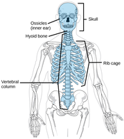
Axial skeleton: Includes the skull, vertebral column, and rib cage.
Appendicular skeleton: Comprises the limbs, shoulder girdle, and pelvic girdle.
Functions of the Skeletal System
The skeletal system performs several essential functions:
Support: Maintains the body’s shape and posture.
Protection: Shields vital organs (e.g., skull protects the brain, rib cage protects thoracic organs).
Movement: Muscles attach to bones, acting as levers for movement.
Storage: Stores minerals (calcium, phosphorus) and fats (yellow marrow).
Hematopoiesis: Formation of blood cells in red marrow.
Classification of Bones
Types of Osseous Tissue
Bones are composed of two main types of tissue:
Compact bone: Dense, smooth, and homogeneous.
Spongy bone: Contains small needlelike pieces (trabeculae) and many open spaces.
Bone Classification by Shape
Bones are classified into four groups based on their shape:
Long bones: Longer than wide, mostly compact bone (e.g., femur, humerus).
Short bones: Cube-shaped, mostly spongy bone (e.g., carpals, tarsals, patella).
Flat bones: Thin, flattened, usually curved, two layers of compact bone sandwiching spongy bone (e.g., skull, ribs, sternum).
Irregular bones: Complex shapes that do not fit other categories (e.g., vertebrae, hip bones).
Tissues of the Skeletal System
Types of Connective Tissue
The skeletal system contains several types of connective tissue:
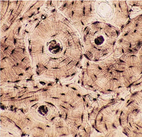
Bone tissue: Provides structural support.
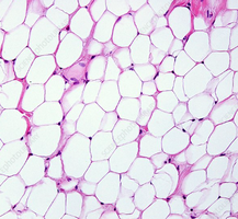
Adipose tissue: Stores fat in yellow marrow.
Hyaline cartilage: Found in joints and rib connections.
Fibrocartilage: Found in intervertebral discs and pubic symphysis.
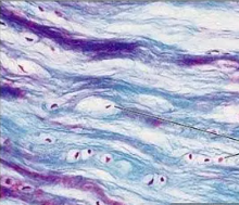
Dense regular connective tissue: Forms ligaments.
Blood tissue: Found in red marrow, responsible for hematopoiesis.
Structure of Bone
Long Bone Anatomy
Long bones have a distinct structure:
Diaphysis: Shaft, made of compact bone.
Epiphysis: Ends, composed mostly of spongy bone.
Periosteum: Fibrous membrane covering the diaphysis.
Articular cartilage: Hyaline cartilage covering epiphyses, reduces friction.
Epiphyseal plate/line: Site of bone growth; plate in youth, line in adults.
Endosteum: Connective tissue lining the inner shaft.
Medullary cavity: Central cavity containing yellow marrow (fat storage) and red marrow (blood cell formation).
Bone Markings
Bone markings are features on bones that serve as sites for muscle, tendon, and ligament attachment, or as passages for nerves and blood vessels.
Projections/processes: Grow out from bone surface.
Depressions/cavities: Indentations in bone.
Microscopic Structure of Bone
Spongy Bone
Spongy bone consists of trabeculae and open spaces filled with marrow, blood vessels, and nerves.
Compact Bone
Compact bone is organized into osteons (Haversian systems):
Osteocytes: Mature bone cells in lacunae.
Lacunae: Cavities housing osteocytes.
Lamellae: Concentric rings around the central canal.
Central (Haversian) canal: Contains blood vessels and nerves.
Canaliculi: Tiny canals connecting lacunae to the central canal.
Perforating (Volkmann’s) canal: Perpendicular canals carrying blood vessels and nerves.
Bone Formation, Growth, and Remodeling
Ossification
Ossification is the process of bone formation, occurring on hyaline cartilage models or fibrous membranes.
Phase 1: Osteoblasts cover cartilage with bone matrix.
Phase 2: Cartilage is replaced by bone matrix, forming the medullary cavity.
Bone Growth
Length: Occurs at articular cartilages and epiphyseal plates; new cartilage forms externally, old cartilage is replaced by bone internally.
Width (Appositional growth): Osteoblasts add bone matrix externally; osteoclasts remove bone internally.
Bone Remodeling
Bone remodeling is a lifelong process influenced by:
Calcium ion levels: Parathyroid hormone (PTH) stimulates osteoclasts when calcium is low; osteoblasts store calcium when levels are high.
Mechanical stress: Pull of gravity and muscles determines where bone is formed or broken down.
Bone Fractures and Repair
Types of Fractures
Closed (simple) fracture: Bone breaks but does not penetrate skin.
Open (compound) fracture: Bone breaks and penetrates skin.
Fracture Treatment and Healing
Reduction: Realignment of bone (closed or open).
Immobilization: Stabilizing bone for healing (6–8 weeks).
Stages of Bone Repair
Hematoma formation
Fibrocartilage callus formation
Bony callus formation
Bone remodeling
Axial Skeleton
Components
The axial skeleton forms the body’s longitudinal axis and includes:
Skull: Cranial and facial bones, joined by sutures.
Vertebral column: Cervical, thoracic, lumbar vertebrae, sacrum, coccyx; separated by fibrocartilage discs.
Bony thorax: Sternum, ribs, thoracic vertebrae.
Appendicular Skeleton
Components
The appendicular skeleton includes:
Limbs: Arms and legs.
Pectoral girdle: Clavicle and scapula.
Pelvic girdle: Coxal bones, sacrum, coccyx.
Joints
Classification of Joints
Joints are classified by function and structure:
Functional: Synarthroses (immovable), amphiarthroses (slightly movable), diarthroses (freely movable).
Structural: Fibrous (immobile), cartilaginous (slightly movable), synovial (freely movable).
Types of Joints
Fibrous joints: United by fibrous tissue (e.g., sutures, syndesmoses, gomphoses).
Cartilaginous joints: Connected by cartilage (e.g., synchondrosis, symphysis).
Synovial joints: Separated by a joint cavity containing synovial fluid; most common in appendages.
Synovial Joint Features
Articular cartilage
Articular capsule
Joint cavity
Reinforcing ligaments
Bursae and tendon sheaths (additional structures)
Types of Synovial Joints
Plane joint: Flat surfaces, gliding movements (e.g., intercarpal joints).
Hinge joint: Uniaxial movement (e.g., elbow, knee).
Pivot joint: Rotation around one axis (e.g., atlas/axis, radioulnar joint).
Condylar joint: Biaxial movement (e.g., metacarpal/phalangeal joints).
Saddle joint: Biaxial movement (e.g., thumb).
Ball-and-socket joint: Multiaxial movement (e.g., shoulder, hip).
Summary Table: Joint Classes
Structural Class | Functional Class | Examples |
|---|---|---|
Fibrous | Synarthroses | Sutures, syndesmoses, gomphoses |
Cartilaginous | Amphiarthroses | Synchondrosis, symphysis |
Synovial | Diarthroses | Plane, hinge, pivot, condylar, saddle, ball-and-socket |
Key Terms and Concepts
Ossification: Process of bone formation.
Osteoblast: Bone-forming cell.
Osteoclast: Bone-destroying cell.
Epiphyseal plate: Growth plate in long bones.
Hematopoiesis: Blood cell formation in red marrow.
Trabeculae: Structural elements in spongy bone.
Lamellae: Concentric rings in compact bone.
Canaliculi: Tiny canals connecting bone cells.
Important Equations
Calcium Regulation:
Bone Growth:
Additional info:
Bone tissue is an organ containing multiple types of connective tissue, including fibrous, cartilage, adipose, and blood.
Bone remodeling is a dynamic process influenced by both hormonal and mechanical factors.
