 뒤로
Study Guide: Ch7CThe Skeleton – Thoracic Cage, Pectoral Girdle, and Upper Limb
스터디 가이드 - 스마트 노트
 자료에 맞춘 맞춤형 노트, 핵심 정의, 예시, 맥락을 확장해 제공합니다.
The Skeleton
The Thoracic Cage
The thoracic cage is a vital bony structure of the chest, composed of the thoracic vertebrae, ribs, sternum, and costal cartilages. It serves to protect essential organs, support the shoulder girdles and upper limbs, and provide attachment sites for muscles.
Components: Thoracic vertebrae (posterior), ribs (lateral), sternum and costal cartilages (anterior)
Functions: Protection of heart and lungs, support for upper limbs, muscle attachment
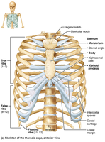
Sternum
The sternum, or breastbone, consists of three fused bones: the manubrium, body, and xiphoid process. It features important anatomical landmarks and is not fully ossified until about age 40.
Manubrium: Superior portion, articulates with clavicular notches and ribs 1 and 2
Body: Midportion, articulates with costal cartilages of ribs 2–7
Xiphoid process: Inferior end, site of muscle attachment
Landmarks: Jugular notch, sternal angle, xiphisternal joint
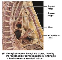
Clinical Note: Xiphoid Process
In some individuals, the xiphoid process projects posteriorly. Trauma to this area can cause severe injury to underlying organs such as the liver or heart. During CPR, hands must be placed on the midsternum to avoid breaking the xiphoid process.
Ribs
The ribs form the sides of the thoracic cage, with 12 pairs attaching posteriorly to the thoracic vertebrae. They are classified as true, false, or floating ribs based on their attachment to the sternum.
True (vertebrosternal) ribs: Pairs 1–7, attach directly to sternum via individual costal cartilages
False (vertebrochondral) ribs: Pairs 8–10, attach indirectly by joining costal cartilage of rib above
Floating (vertebral) ribs: Pairs 11–12, no attachment to sternum

Structure of a Typical Rib
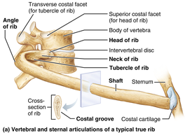
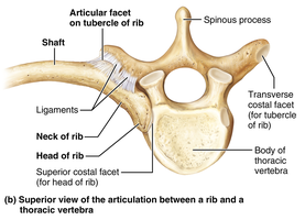
Each rib has several distinct parts that facilitate articulation and muscle attachment.
Shaft: Flat bone forming most of the rib
Head: Posterior end, articulates with vertebrae
Neck: Constricted portion beyond head
Tubercle: Knoblike structure, articulates with transverse costal facet
The Appendicular Skeleton
Pectoral Girdle
The pectoral girdle consists of the clavicle and scapula, attaching the upper limbs to the body trunk. Its structure allows for a high degree of mobility and provides muscle attachment sites.
Clavicle: S-shaped bone, articulates medially with sternum and laterally with scapula
Scapula: Thin, triangular bone on dorsal surface of rib cage
Mobility: Scapulae are not attached to axial skeleton; shoulder joint socket is shallow
Clavicle
Sternal end: Articulates with sternum
Acromial end: Articulates with scapula
Function: Anchors muscles, acts as brace for scapulae and arms
Scapula
Borders: Superior, medial (vertebral), lateral (axillary)
Angles: Superior, lateral, inferior
Features: Spine, acromion, coracoid process, suprascapular notch, fossae (subscapular, supraspinous, infraspinous)
The Upper Limb
Arm
The arm contains the humerus, the largest and longest bone of the upper limb. It articulates with the scapula, radius, and ulna.
Head: Fits into glenoid cavity of scapula
Anatomical neck: Slight constriction below head
Greater and lesser tubercles: Sites of rotator cuff muscle attachment
Surgical neck: Most frequently fractured part
Radial groove: Carries radial nerve
Trochlea and capitulum: Distal condyles for articulation
Epicondyles: Muscle attachment points
Fossae: Coronoid, olecranon, radial
Forearm
The forearm consists of two parallel bones: the ulna (medial) and radius (lateral). They articulate with the humerus and each other, connected by the interosseous membrane.
Ulna: Forms major portion of elbow joint; features olecranon and coronoid processes, trochlear notch, radial notch, ulnar head, and styloid process
Radius: Articulates with capitulum of humerus and radial notch of ulna; features head, radial tuberosity, ulnar notch, and styloid process
Clinical Note: Colles’ Fracture
A break in the distal end of the radius, common when a person falls and attempts to break the fall with outstretched hands.
Hand
The hand is composed of the carpus (wrist), metacarpus (palm), and phalanges (fingers).
Carpus: Eight bones in two rows (proximal: scaphoid, lunate, triquetrum, pisiform; distal: trapezium, trapezoid, capitate, hamate)
Metacarpus: Five metacarpal bones (I–V) from thumb to little finger
Phalanges: Fingers I–V; thumb (pollex) has two bones, other fingers have three (distal, middle, proximal)
Clinical Note: Carpal Tunnel Syndrome
The median nerve and tendons pass through the carpal tunnel, formed by ligaments. Overuse and inflammation can compress the median nerve, causing tingling and numbness.
Table: Bones of the Appendicular Skeleton (Pectoral Girdle and Upper Limb)
Region | Bones | Key Features |
|---|---|---|
Pectoral Girdle | Clavicle, Scapula | Mobility, muscle attachment |
Arm | Humerus | Head, tubercles, neck, epicondyles |
Forearm | Ulna, Radius | Olecranon, coronoid, radial tuberosity, styloid processes |
Hand | Carpus, Metacarpus, Phalanges | Wrist bones, palm, fingers |
Additional info: The notes above expand on brief points with academic context, definitions, and clinical relevance to ensure completeness and clarity for exam preparation.
