 뒤로
Study Guide: The Spinal Cord, Spinal Nerves, and Spinal Reflexes
스터디 가이드 - 스마트 노트
 자료에 맞춘 맞춤형 노트, 핵심 정의, 예시, 맥락을 확장해 제공합니다.
The Nervous System: Structural Organization and Reflexes
Central and Peripheral Nervous System
The nervous system is divided into the central nervous system (CNS) and peripheral nervous system (PNS). The CNS consists of the brain and spinal cord, serving as processing centers for sensory input and motor output. The PNS includes cranial and spinal nerves, which transmit sensory information to the CNS and carry motor commands to effectors.
Reflexes: Quick, automatic responses to specific stimuli.
Spinal reflexes: Controlled by the spinal cord alone, independent of brain input.
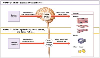
The Spinal Cord: Anatomy and Function
Gross Anatomy of the Spinal Cord
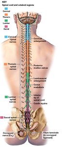
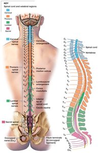
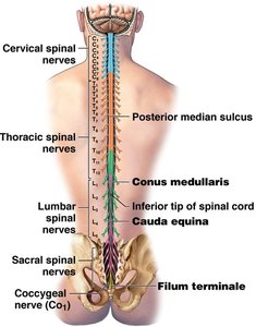
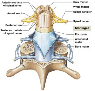
The spinal cord is a nervous tissue organ housed within protective membranes (meninges) and the vertebral column. It carries sensory and motor information between the brain and most body parts and is the origin of spinal nerves.
Length: ~18 inches (45 cm); width: ~0.55 inches (14 mm).
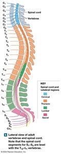
Extends from the brain to vertebrae L1 and L2.
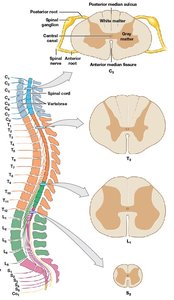
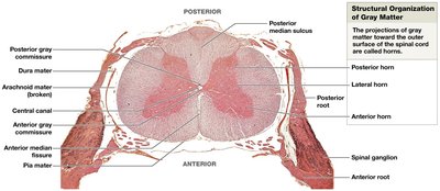
Contains gray matter (neuronal cell bodies) and white matter (myelinated axons).
Four regions: cervical, thoracic, lumbar, sacral.
31 segments, each giving rise to a pair of spinal nerves.
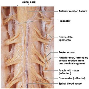
Bilateral symmetry; divided by posterior median sulcus and anterior median fissure.
Central canal: Contains cerebrospinal fluid (CSF).
Enlargements: Cervical (shoulder/upper limb) and lumbosacral (pelvis/lower limb).
Spinal Roots and Ganglia
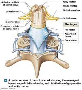
Spinal nerves attach to the spinal cord via two roots:
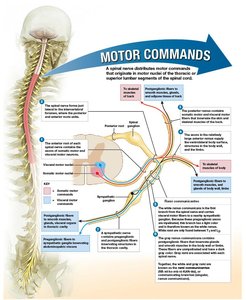
Anterior (ventral) root: Motor neuron axons.
Posterior (dorsal) root: Sensory neuron axons.
Roots divide into rootlets before entering/leaving the cord.
Spinal ganglia (dorsal root ganglia): Contain cell bodies of sensory neurons; located between vertebral pedicles.
Spinal Nerves and Branches
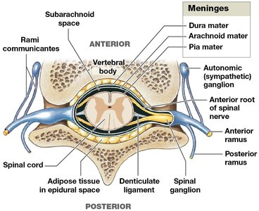
Spinal nerves are formed by the union of anterior and posterior roots and are mixed nerves (contain both sensory and motor fibers). They branch into:
White ramus communicans: Myelinated axons to glands/smooth muscle.
Gray ramus communicans: Unmyelinated fibers to glands/smooth muscle.
Posterior ramus: Innervates skin/muscles of the back.
Anterior ramus: Innervates ventrolateral trunk and limbs.
Naming Spinal Nerves
Named by vertebral region and number (e.g., C1 above first cervical vertebra, C8 below seventh cervical vertebra).
All others named for the vertebra above.
Spinal Meninges: Structure and Function
Three Layers of Meninges
The spinal cord is surrounded by three specialized membranes:
Dura mater: Tough, outer fibrous layer; continuous with cranial dura mater; forms coccygeal ligament with filum terminale.
Arachnoid mater: Middle layer; includes arachnoid membrane and trabeculae; subarachnoid space filled with CSF.
Pia mater: Innermost layer; mesh of collagen/elastic fibers; firmly attached to neural tissue; blood vessels on surface; denticulate ligaments anchor pia to other meninges.
Epidural space: Between vertebrae and dura mater; contains loose connective and adipose tissue; site for epidural anesthesia.
Subarachnoid space: Between arachnoid and pia mater; filled with CSF; site for lumbar puncture/spinal tap.
Meningitis: Infection of meninges (viral or bacterial).
Gray Matter and White Matter of the Spinal Cord
Gray Matter: Structure and Function
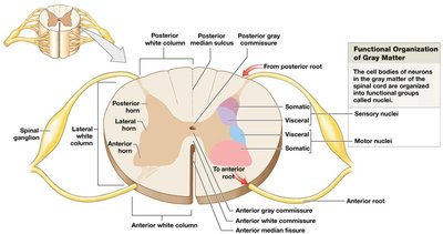
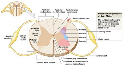
Gray matter consists of neuron cell bodies, neuroglia, and unmyelinated axons. It integrates information and initiates commands, located deep within the spinal cord.
Nuclei: Masses of gray matter in CNS, organized into horns.
Posterior horns: Somatic and visceral sensory nuclei.
Anterior horns: Somatic motor nuclei.
Lateral horns: Visceral motor nuclei (thoracic/lumbar segments).
Gray commissure: Narrow band around central canal.
White Matter: Structure and Function
White matter consists of myelinated and unmyelinated axons, carrying information between regions. It is superficial in the spinal cord and organized into columns:
Posterior white columns: Between posterior horns and posterior median sulcus.
Anterior white columns: Between anterior horns and anterior median fissure; connected by anterior white commissure.
Lateral white columns: On each side between anterior and posterior columns.
Tracts: Bundles of axons relaying same type of information in same direction.
Ascending tracts: Sensory information toward the brain.
Descending tracts: Motor commands down to the spinal cord.
Spinal Nerves and Plexuses
Connective Tissue Layers of Spinal Nerves
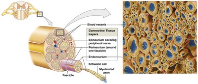
Spinal nerves are surrounded by three connective tissue layers:
Epineurium: Outermost, network of collagen fibers.
Perineurium: Middle, separates nerve into fascicles (bundles of axons).
Endoneurium: Innermost, surrounds individual axons.
Peripheral Distribution and Dermatome
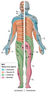
Each spinal cord segment gives rise to a pair of spinal nerves.
Dermatome: Specific bilateral region of skin monitored by a single pair of spinal nerves.
Peripheral neuropathies: Regional loss of sensory/motor function due to trauma or compression.
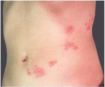
Shingles: Varicella-zoster virus attacks spinal nerves, causing painful rash along dermatomes.
Nerve Plexuses
A nerve plexus is a complex, interwoven network of nerve fibers formed from blended fibers of the anterior rami of adjacent spinal nerves. Plexuses allow multiple spinal nerves to supply the same structures.
Four major plexuses: cervical, brachial, lumbar, sacral.
Cervical Plexus
Anterior rami of C1–C5.
Innervates scalp behind ear, neck, diaphragm.
Major nerves: phrenic (diaphragm), lesser occipital, great auricular, transverse cervical, supraclaviculars, ansa cervicalis.
Brachial Plexus
Anterior rami of C5–T1.
Innervates pectoral girdle, upper back, upper limbs.
Trunks (superior, middle, inferior) form cords (lateral, posterior, medial).
Major nerves: musculocutaneous, radial, median, ulnar.
Lumbar Plexus
Anterior rami of T12–L4.
Major nerves: iliohypogastric, ilio-inguinal, genitofemoral, lateral femoral cutaneous, femoral, obturator.
Sacral Plexus
Anterior rami of L4–S4.
Major nerves: superior gluteal, inferior gluteal, posterior femoral cutaneous, sciatic (fibular and tibial branches), pudendal.
Neuronal Pools and Circuit Patterns
Functional Organization of Neurons
Neurons are organized into functional groups called neuronal pools, which have limited input sources and output destinations. They may stimulate or depress parts of the CNS.
Sensory neurons: Bring information to CNS.
Motor neurons: Send commands from CNS to effectors.
Interneurons: Interpret, plan, and coordinate signals within CNS.
Patterns of Neural Circuits
Divergence: Spreads information from one neuron to many; common in sensory pathways.
Convergence: Several neurons synapse on a single neuron; e.g., control of diaphragm.
Serial processing: Information moves sequentially through neurons; e.g., pain signals.
Parallel processing: Several neurons process same information simultaneously; e.g., response to stepping on a sharp object.
Reverberation: Collateral branches stimulate presynaptic neurons, forming positive feedback loops; e.g., maintaining consciousness, breathing, muscle coordination.
Summary Table: Spinal Cord Regions and Functions
Region | Major Function | Associated Structures |
|---|---|---|
Cervical | Upper limb and neck innervation | Cervical enlargement, phrenic nerve |
Thoracic | Trunk and upper back | Intercostal nerves |
Lumbar | Lower limb and pelvis | Lumbosacral enlargement, femoral nerve |
Sacral | Lower limb, perineum | Sciatic nerve, pudendal nerve |
Key Terms and Definitions
Reflex: Automatic response to a stimulus.
Spinal nerve: Mixed nerve emerging from spinal cord segment.
Dermatome: Skin region innervated by a specific spinal nerve.
Plexus: Network of interwoven nerve fibers.
Meninges: Protective membranes surrounding CNS.
Gray matter: Neuron cell bodies and unmyelinated axons.
White matter: Myelinated axons transmitting information.
