 뒤로
Study Guide: The Vertebral Column and Regional Vertebral Characteristics
스터디 가이드 - 스마트 노트
 자료에 맞춘 맞춤형 노트, 핵심 정의, 예시, 맥락을 확장해 제공합니다.
The Vertebral Column: Structure and Function
General Characteristics
The vertebral column, also known as the spine or spinal column, is a flexible, curved structure that extends from the skull to the pelvis. It serves several essential functions in the human body:
Transmit weight of the trunk to the lower limbs
Surround and protect the spinal cord
Provide attachment points for ribs and muscles
The adult vertebral column contains 26 irregular bones called vertebrae, organized into five major regions. In infants, there are 33 vertebrae, some of which fuse during development.
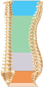
Regions of the Vertebral Column
The vertebral column is approximately 28 inches long and is divided into five major regions:
Cervical: 7 vertebrae (C1–C7)
Thoracic: 12 vertebrae (T1–T12)
Lumbar: 5 vertebrae (L1–L5)
Sacrum: 1 bone formed from the fusion of 5 vertebrae; articulates with the hip
Coccyx: 4 fused bones; forms the terminus of the column
Mnemonic: Remember meal times—7 am (cervical), 12 noon (thoracic), and 5 pm (lumbar).

Curvatures of the Vertebral Column
The vertebral column has four main curvatures that increase its resilience and flexibility:
Cervical and lumbar curvatures: Concave posteriorly
Thoracic and sacral curvatures: Convex posteriorly
These curvatures help distribute mechanical stress during movement and standing.

Clinical Considerations: Abnormal Spinal Curvatures
Types of Abnormal Curvatures
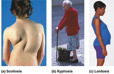
Abnormal spinal curvatures may be congenital or result from disease, poor posture, or unequal muscle pull. The main types include:
Scoliosis: Abnormal lateral rotation of the spine, most often in the thoracic region; may cause breathing difficulties.
Kyphosis (hunchback): Abnormal dorsal thoracic curvature; common in osteoporosis, tuberculosis, rickets, or osteomalacia.
Lordosis (swayback): Accentuation of lumbar curvature; seen in disease, males with pot bellies, and pregnant females.
Ligaments and Intervertebral Discs
Ligaments Supporting the Vertebral Column
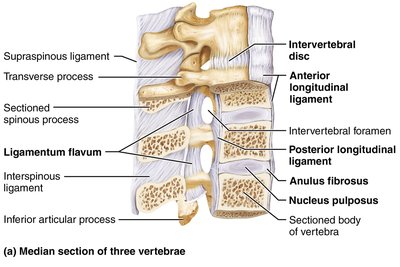
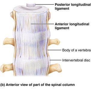
Ligaments, along with trunk muscles, help support the vertebral column:
Anterior and posterior longitudinal ligaments: Continuous bands from neck to sacrum; prevent hyperextension and hyperflexion.
Ligamentum flavum: Connects adjacent vertebrae.
Short ligaments: Connect each vertebra to those above and below.
Intervertebral Discs
Intervertebral discs are cushion-like pads between vertebrae that act as shock absorbers. Each disc consists of two parts:
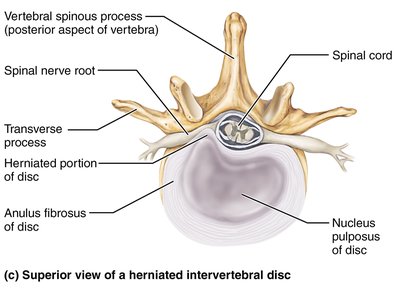
Nucleus pulposus: Inner gelatinous nucleus; provides elasticity and compressibility.
Anulus fibrosus: Outer collar of collagen and fibrocartilage; limits expansion of nucleus pulposus when compressed.
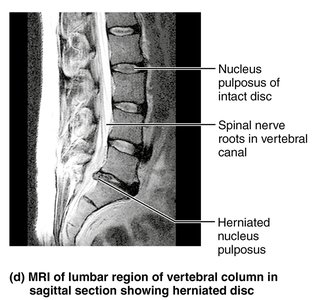
Clinical: Herniated (Prolapsed) Discs
Severe trauma may cause rupture of the anulus fibrosus, resulting in protrusion of the nucleus pulposus. This can press on the spinal cord or nerves, causing pain or numbness. Treatment options include exercise, massage, heat, painkillers, or surgery.

Structure of a Typical Vertebra
General Structure
All vertebrae share a common structural pattern:
Body: Anterior weight-bearing region
Vertebral arch: Composed of two pedicles (sides) and two laminae (posterior plates)
Vertebral foramen: Enclosure formed by body and arch
Vertebral canal: Series of vertebral foramina
Intervertebral foramina: Lateral openings for spinal nerves
Processes of Vertebrae
Each vertebra has seven processes:
Spinous process: Projects posteriorly
Transverse processes (2): Project laterally
Superior articular processes (2): Protrude superiorly
Inferior articular processes (2): Protrude inferiorly
Regional Vertebral Characteristics
Cervical Vertebrae (C1–C7)
The cervical vertebrae are the smallest and lightest. Key features include:
Oval-shaped body
Bifid spinous processes (except C7)
Large, triangular vertebral foramen
Transverse foramen in each transverse process for artery passageways
C7 is vertebra prominens, palpable through the skin
Atlas (C1): No body or spinous process; articulates with occipital condyles for nodding "Yes".
Axis (C2): Has a knoblike dens projecting superiorly; allows rotation for saying "No".
Thoracic Vertebrae (T1–T12)
Thoracic vertebrae increase in size and articulate with ribs. Unique features:
Heart-shaped body with two small demifacets for rib articulation
Circular vertebral foramen
Long, sharp spinous process pointing inferiorly
Transverse costal facets for rib articulation (except T11, T12)
Articular facet orientation allows rotation
Lumbar Vertebrae (L1–L5)
The lumbar vertebrae are located in the "small of the back" and receive the most stress. Features include:
Massive, kidney-shaped bodies
Short, thick pedicles and laminae
Flat, hatchet-shaped spinous processes pointing posteriorly
Triangular vertebral foramen
Articular facet orientation locks vertebrae together, preventing rotation
Comparison Table: Regional Characteristics of Cervical, Thoracic, and Lumbar Vertebrae
Characteristic | Cervical (3–7) | Thoracic | Lumbar |
|---|---|---|---|
Body | Small, oval, wide side to side | Heart-shaped; bears two costal facets | Massive; kidney-shaped |
Spinous process | Short; bifid (except C7); projects directly posterior | Long; sharp; projects inferiorly | Short; blunt; rectangular; projects directly posteriorly |
Vertebral foramen | Triangular, large | Circular | Triangular |
Transverse processes | Contain foramina | Bear facets for ribs (except T11, T12) | Thin and tapered |
Superior and inferior articular processes | Superior facets directed superoposteriorly; Inferior facets inferoanteriorly | Superior facets directed posteriorly; Inferior facets anteriorly | Superior facets posteromedially; Inferior facets anterolaterally |
Movements allowed | Flexion, extension, lateral flexion, rotation; greatest range of movement | Rotation, lateral flexion (restricted by ribs), limited flexion/extension | Flexion, extension, some lateral flexion; rotation prevented |
Sacrum and Coccyx
Sacrum
The sacrum is a triangular bone forming the posterior wall of the pelvis, made from five fused vertebrae (S1–S5). It articulates with the coccyx inferiorly and hip bones laterally via auricular surfaces, forming sacroiliac joints. Key features:
Sacral promontory: Anterosuperior margin
Transverse ridges: Lines of fusion
Anterior and posterior sacral foramina: Openings for nerves and vessels
Alae: Winglike expansions
Median and lateral sacral crests: Roughened areas on posterior side
Sacral canal: Continuation of vertebral canal
Sacral hiatus: Large opening at end of canal
Coccyx
The coccyx, or tailbone, is formed from three to five fused vertebrae and articulates superiorly with the sacrum. It has very little function in humans.
