 뒤로
Study Notes: The Central Nervous System (CNS) - Structure and Function
스터디 가이드 - 스마트 노트
 자료에 맞춘 맞춤형 노트, 핵심 정의, 예시, 맥락을 확장해 제공합니다.
The Central Nervous System
Overview and Directional Terms
The Central Nervous System (CNS) consists of the brain and spinal cord, serving as the main control center for the body. Unique directional terms are used in CNS anatomy:
Rostral: Toward the nose (anterior)
Caudal: Toward the tail (posterior)
Functions of the Brain
The brain regulates vital functions such as heart rate, respiratory rate, and blood pressure. It controls the internal environment via the autonomic nervous system and endocrine system, and innervates the head through cranial nerves. The brain is responsible for complex neural functions including intelligence, consciousness, memory, sensory-motor integration, emotion, behavior, and socialization.
Basic Parts and Organization of the Brain
Major Regions
The brain is classified into four main regions:
Brain Stem: Includes the midbrain, pons, and medulla oblongata
Cerebellum
Diencephalon
Cerebrum: Composed of two cerebral hemispheres
Ventricles of the Brain
The brain contains four interconnected ventricles, which are expansions of the central cavity filled with cerebrospinal fluid (CSF) and lined with ependymal cells. These ventricles are continuous with each other and with the central canal of the spinal cord.
Lateral Ventricles: Located in the cerebral hemispheres, horseshoe-shaped
Third Ventricle: Located in the diencephalon, connected to lateral ventricles by the interventricular foramen
Cerebral Aqueduct: Connects the third and fourth ventricles
Fourth Ventricle: Located at the junction of the cerebellum and brain stem, connects to the central canal of the spinal cord
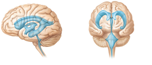
Distribution of Gray and White Matter
The CNS exhibits a distinct pattern of gray and white matter:
Gray Matter: Contains neuronal cell bodies and short nonmyelinated neurons; centrally located in the spinal cord, forms the cortex in the cerebrum and cerebellum
White Matter: Contains mostly myelinated axons; located externally to gray matter
In the cerebrum and cerebellum, an additional layer of gray matter (the cortex) is found external to the white matter.
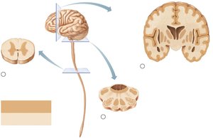
The Brain Stem
Structure and Functions
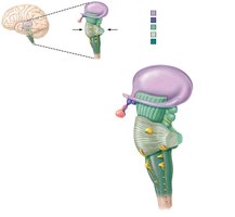
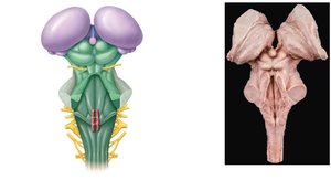
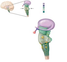
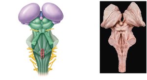
The brain stem consists of the midbrain, pons, and medulla oblongata. It serves as a passageway for all fiber tracts running between the cerebrum and spinal cord, is involved in the innervation of the face and head (10 of 12 cranial nerves attach here), produces automatic behaviors necessary for survival, and integrates auditory and visual reflexes.
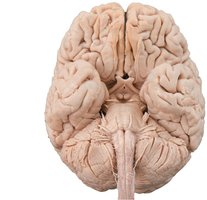
Medulla Oblongata
The medulla oblongata is the most caudal part of the brain stem and is continuous with the spinal cord. It contains major fiber tracts and nuclei, including:
Pyramids: Motor tracts on the ventral surface
Decussation of the pyramids: Crossing over of motor tracts
Inferior cerebellar peduncles: Connect medulla and cerebellum
Olive: Contains the inferior olivary nucleus, a relay for proprioceptive information
The core of the medulla contains much of the reticular formation, which influences autonomic functions such as cardiac, vasomotor, and respiratory centers.
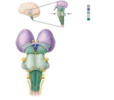
Pons
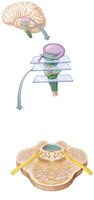
The pons acts as a bridge between the midbrain and medulla oblongata. It contains nuclei of cranial nerves and motor tracts from the cerebral cortex. Pontine nuclei connect the cerebral cortex and cerebellum, sending axons to the cerebellum via the middle cerebellar peduncles.
Midbrain
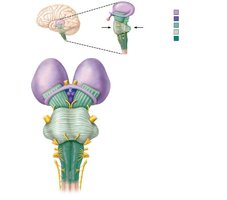
The midbrain lies between the diencephalon and pons. It contains the cerebral aqueduct, cerebral peduncles (pyramidal tracts), and superior cerebellar peduncles. The periaqueductal gray matter surrounds the aqueduct and is involved in fight-or-flight reactions and visceral pain responses. The corpora quadrigemina (superior and inferior colliculi) are nuclei involved in visual and auditory reflexes. The substantia nigra and red nucleus are involved in motor control.
The Cerebellum
Structure and Function
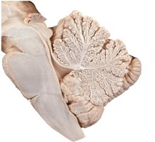
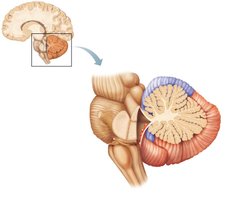
The cerebellum is located dorsal to the pons and medulla. It smooths and coordinates body movements and helps maintain equilibrium. The surface is folded into ridges called folia, and the hemispheres are subdivided into anterior, posterior, and flocculonodular lobes.
Anterior lobe: Coordinates trunk and limb movements
Posterior lobe: Participates in cognitive and emotional functions
Flocculonodular lobe: Adjusts posture and coordinates head/eye movements
Cerebellar Peduncles
Thick tracts connect the cerebellum to the brain stem:
Superior cerebellar peduncles
Middle cerebellar peduncles
Inferior cerebellar peduncles
Fibers to and from the cerebellum are ipsilateral (same side).
The Diencephalon
Structure and Components
The diencephalon forms the central core of the forebrain and is composed of three paired structures:
Thalamus
Hypothalamus
Epithalamus
These border the third ventricle and are primarily composed of gray matter.
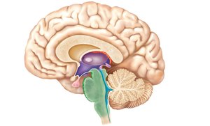
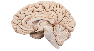
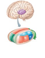
Thalamus
The thalamus makes up 80% of the diencephalon and acts as a relay station for incoming sensory messages. It contains a dozen major nuclei, which organize, amplify, or tone down signals before sending them to the cerebral cortex.
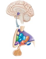
Hypothalamus
The hypothalamus is the main visceral control center of the body, regulating the autonomic nervous system, emotional responses, body temperature, hunger, thirst, behavior, sleep-wake cycles, endocrine system, and memory formation.
Epithalamus
The epithalamus forms part of the roof of the third ventricle and includes the pineal gland, which secretes melatonin and helps regulate circadian rhythms.
The Cerebrum
Structure and Surface Features
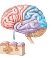
The cerebrum accounts for 83% of brain mass and is divided into two hemispheres. Deep grooves called fissures separate major regions, while sulci (grooves) and gyri (ridges) mark the surface. Prominent gyri and sulci are similar in all people.
Transverse fissure: Separates cerebrum and cerebellum
Longitudinal fissure: Separates cerebral hemispheres
Central sulcus: Separates frontal and parietal lobes
Lobes of the Cerebral Cortex
The cerebrum is divided into lobes named for the overlying skull bones:
Frontal lobe
Parietal lobe
Occipital lobe
Temporal lobe
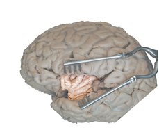
Insula: Deep within the lateral sulcus
Cerebral Cortex
The cerebral cortex is the home of our conscious mind, enabling awareness, voluntary movement, communication, memory, and understanding. It is composed of gray matter and is highly folded, tripling its surface area. The cortex is divided into 47 Brodmann areas, each with distinct functions.
Functional Areas of the Cerebral Cortex
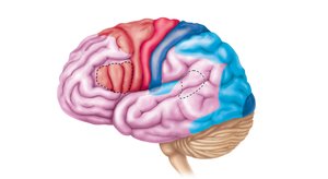
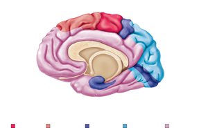
Three general kinds of functional areas exist:
Sensory areas: Receive and process sensory information
Association areas: Integrate and interpret sensory input
Motor areas: Plan and initiate voluntary movements
Sensory Areas
Sensory areas are located in the parietal, temporal, and occipital lobes. Each major sense has a primary sensory cortex and an association area for processing.
Primary Somatosensory Cortex
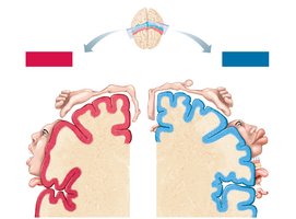
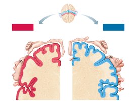
Located along the postcentral gyrus, this area is involved in conscious awareness of general somatic senses and spatial discrimination. The sensory homunculus is a body map of the sensory cortex, with contralateral projection (each hemisphere receives input from the opposite side of the body).
Somatosensory Association Cortex
Lies posterior to the primary somatosensory cortex and integrates different sensory inputs, allowing recognition of objects by touch.
Visual, Auditory, Vestibular, Gustatory, and Olfactory Areas
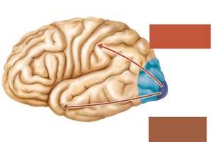
Primary visual cortex: Deep within the calcarine sulcus, receives visual information
Visual association area: Surrounds primary visual cortex, analyzes color, form, and movement
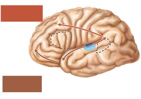
Primary auditory cortex: Superior edge of temporal lobe, conscious awareness of sound
Auditory association area: Permits evaluation of different sounds
Vestibular cortex: Posterior part of insula, awareness of balance
Gustatory cortex: In insula, awareness of taste
Olfactory cortex: Medial aspect of cerebrum, awareness of smells
Motor Areas
Motor areas are localized in the posterior frontal lobe and include the premotor cortex, primary motor cortex, frontal eye field, and Broca’s area. The primary motor cortex (precentral gyrus) contains pyramidal cells that control specific areas of the body, with somatotopy (spatial representation).
Multimodal Association Areas
These areas receive input from multiple sensory modalities and association areas, integrating information for complex functions such as perception, memory, and planning. The three main multimodal association areas are:
Posterior association area: Integrates sensory information, spatial awareness, language comprehension
Anterior association area: Prefrontal cortex, involved in thinking, planning, judgment, and social skills
Limbic association area: Memory and emotions
Visual and Auditory Pathways
Visual and auditory information is processed in dorsal ("where") and ventral ("what") streams, allowing spatial localization and identification of objects and sounds.
Lateralization of Cortical Functioning
The two cerebral hemispheres control opposite sides of the body and are specialized for different cognitive functions. The left hemisphere is dominant for language, math, and logic, while the right hemisphere is involved in visual-spatial skills, emotion, and artistic abilities.
Cerebral White Matter
Types of Fiber Tracts
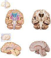
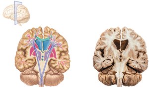
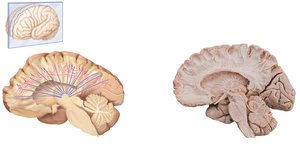
White matter consists of myelinated axons bundled into tracts:
Commissural fibers: Connect the two hemispheres (e.g., corpus callosum)
Association fibers: Connect different parts of the same hemisphere
Projection fibers: Run vertically, connecting the cortex with lower regions
Deep Gray Matter of the Cerebrum
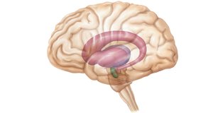
Basal Nuclei (Ganglia)
The basal nuclei are deep within the cerebral white matter and include the caudate nucleus, putamen, and globus pallidus. They cooperate with the cerebral cortex in controlling movement, starting, stopping, and regulating the intensity of voluntary movements.
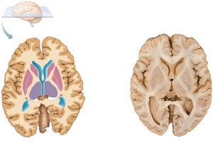
Basal Forebrain Nuclei
These nuclei synthesize and release acetylcholine and are involved in arousal, learning, memory, and motor control. Degeneration is associated with Alzheimer’s disease.
Functional Brain Systems
Limbic System
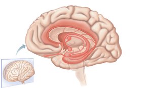
The limbic system is the "emotional brain," involved in emotion, memory, and visceral responses. It includes the cingulate gyrus, hippocampal formation, amygdaloid body, and is linked by the fornix and other tracts.
Reticular Formation
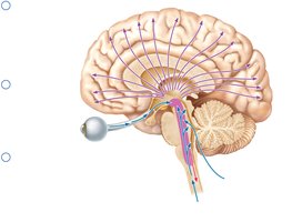
The reticular formation runs through the brain stem and is ideal for arousal of the brain as a whole. The reticular activating system (RAS) maintains consciousness and alertness, and functions in sleep and arousal.
Protection of the Brain
Meninges
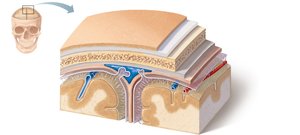
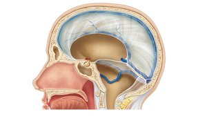
The brain is protected by the skull, meninges, cerebrospinal fluid, and the blood-brain barrier. The meninges include:
Dura mater: Strongest, two layers (periosteal and meningeal)
Arachnoid mater: Beneath dura, contains arachnoid villi for CSF absorption
Pia mater: Delicate, clings tightly to the brain surface
Cerebrospinal Fluid (CSF)
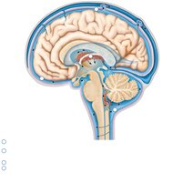
CSF fills the cavities of the brain and spinal cord, providing a cushion, nourishment, waste removal, and chemical signaling. It is formed in choroid plexuses in the ventricles and circulates through the CNS.
Blood-Brain Barrier
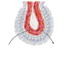
The blood-brain barrier prevents most blood-borne toxins from entering the brain, but allows passage of nutrients, alcohol, nicotine, and anesthetics.
The Spinal Cord
Structure and Function
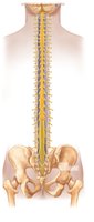
The spinal cord provides a two-way conduction pathway and is a major center for reflexes. It runs through the vertebral canal and extends from the foramen magnum to the level of the vertebra. Key features include the conus medullaris, filum terminale, cervical and lumbar enlargements, and cauda equina.
Spinal Cord Segments
Spinal cord segments are designated by the spinal nerve that issues from them. In lower regions, segments are not adjacent to their corresponding vertebrae due to differential growth rates during development.
White and Gray Matter of the Spinal Cord
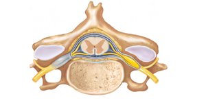
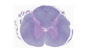
The spinal cord has an outer region of white matter (myelinated and nonmyelinated axons) and an inner region of gray matter (neuronal cell bodies). Gray matter is shaped like an "H" and divided into dorsal, ventral, and lateral horns.
Organization of Gray Matter
Gray matter is divided according to somatic and visceral regions:
SS: Somatic sensory
VS: Visceral sensory
VM: Visceral motor
SM: Somatic motor
Protection of the Spinal Cord
The spinal cord is protected by vertebrae, meninges, and CSF. The meninges include a single layer of dura mater, arachnoid mater, and pia mater, which extends to the coccyx and forms denticulate ligaments.
Sensory and Motor Pathways in the CNS
Multineuron pathways connect the brain and body periphery. Ascending pathways carry information to more rostral areas, while descending pathways carry information to more caudal regions. Ascending pathways include the dorsal column, spinothalamic, and spinocerebellar tracts. Descending pathways include direct (pyramidal) and indirect motor pathways.
Region | Function |
|---|---|
Medulla oblongata | Contains projection fibers, decussation of pyramids, relays sensory and motor information, controls autonomic functions |
Pons | Contains projection fibers, pontine nuclei, connects cortex and cerebellum |
Midbrain | Contains cerebral peduncles, superior/inferior colliculi, substantia nigra, red nucleus |
Cerebellum | Coordinates movement, maintains equilibrium, involved in cognition |
Thalamus | Relay station for sensory information |
Hypothalamus | Controls autonomic and endocrine functions, regulates homeostasis |
Cerebral cortex | Conscious mind, sensory/motor integration, memory, language |
Basal nuclei | Motor control, movement regulation |
Limbic system | Emotion, memory |
Example: The decussation of pyramids in the medulla oblongata explains why each hemisphere of the brain controls the opposite side of the body.
Additional info: The notes above expand on brief points with academic context, definitions, and examples to ensure completeness and clarity for exam preparation.
