 뒤로
Study Notes: The Muscular System – Structure, Function, and Physiology
스터디 가이드 - 스마트 노트
 자료에 맞춘 맞춤형 노트, 핵심 정의, 예시, 맥락을 확장해 제공합니다.
The Muscular System
Muscle Twitch and Measurement
A muscle twitch is a brief contraction of a muscle fiber in response to a single stimulus. It is primarily a laboratory phenomenon used to study tension production in muscle fibers. The twitch is measured using a myogram, which records the tension produced by a muscle fiber over time after stimulation by an electrode that simulates an action potential.
Latent period: Time between stimulus and onset of contraction; action potential spreads through sarcoplasm.
Contraction period: Tension increases as crossbridge cycling occurs.
Relaxation period: Tension decreases as Ca2+ is pumped back into the sarcoplasmic reticulum (SR).
Refractory period: Time during which the muscle cannot respond to another stimulus.
Muscle Tension from Twitches
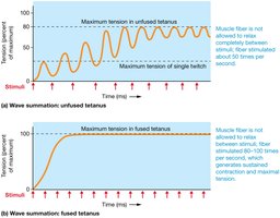
The amount of tension a myofibril can produce depends on several factors, including the frequency of stimuli and the number of crossbridges formed. Wave summation occurs when stimuli are delivered in rapid succession, leading to increased tension. There are two types:
Unfused tetanus: Muscle fiber is not allowed to relax completely between stimuli, resulting in fluctuating tension.
Fused tetanus: Muscle fiber is stimulated so rapidly that no relaxation occurs, resulting in sustained maximal tension.
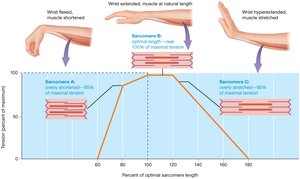
The length-tension relationship describes how the overlap between actin and myosin affects tension production. Optimal overlap allows for maximal crossbridge formation and tension.
Variation in Muscle Fibers
Muscle fibers are classified based on their contraction speed and metabolic properties:
Type I fibers (slow twitch, "dark meat"): Low myosin ATPase activity, maintain extended periods of contraction, rely on oxidative metabolism, have abundant blood supply, myoglobin, and mitochondria. Also called slow oxidative fibers.
Type II fibers (fast twitch, "white meat"): High myosin ATPase activity, quickly fatigued but powerful, rely on anaerobic metabolism, have less blood supply, myoglobin, and mitochondria. Subtypes include fast oxidative glycolytic fibers, fast oxidative fibers, and fast glycolytic fibers (fastest).
Motor Units and Muscle Fiber Arrangement
A motor unit consists of a single motor neuron and all the muscle fibers it innervates. Motor units vary in size depending on the muscle's function:
Large muscles (e.g., gastrocnemius): 2000–3000 fibers per motor unit.
Small muscles (e.g., finger muscles): ~10 fibers per motor unit.
All fibers within a motor unit are of the same type (fast or slow).
Multiple motor units exist within a single muscle.
Muscle fibers are arranged functionally into motor units, which are recruited as needed to produce varying levels of force. Recruitment refers to the activation of additional motor units to increase muscle tension. Muscle tone is maintained by alternating motor unit contractions, and shivering involves random motor unit contractions to generate heat.
Types of Muscle Contraction
Muscle contractions are classified based on changes in muscle length and tension:
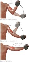
Isotonic concentric: Muscle shortens as force generated exceeds the load.
Isotonic eccentric: Muscle lengthens as force generated is less than the load.
Isometric: Muscle length does not change, but tension increases.
The Muscular System (II)
Fascicle Arrangement and Muscle Shapes
Muscles are organized into fascicles, which are bundles of muscle fibers. The arrangement of fascicles determines the shape and function of the muscle:
Parallel: Sartorius
Convergent: Pectoralis major
Unipennate: Flexor pollicis longus
Bipennate: Rectus femoris
Multipennate: Deltoid
Circular (sphincter): Orbicularis oculi
Spiral: Supinator
Fusiform: Biceps brachii
Muscle Naming Conventions
Muscles are named based on several criteria:
Shape/appearance: Deltoid (triangular), Gastrocnemius (stomach-shaped)
Size: Adductor longus (long), Vastus lateralis (wide)
Number of heads: Biceps (2 heads), Triceps (3 heads), Quadriceps (4 heads)
Points of attachment: Sternocleidomastoid
Location: Tibialis anterior, Tibialis posterior
Body region: Biceps brachii, Biceps femoris
Action: Abductor pollicis longus (abduction)
Functions of Skeletal Muscles
Skeletal muscles perform a variety of functions essential for movement and homeostasis:
Locomotion: Movement across articulations
Breathing: Diaphragm and intercostals
Swallowing initiation
Sound modulation: Stapedius in middle ear
Voluntary control over defecation and urination
Shivering: Heat generation
Muscles work in functional groups:
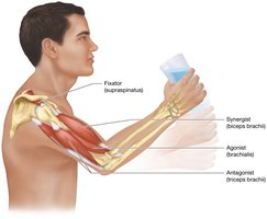
Agonists (prime movers): Provide most force for a given action
Antagonists: Oppose the action of agonists, allowing modulation and control
Synergists: Aid agonists by supplying supplemental force and stabilizing joints
Fixators: Anchor bones, providing movement efficiency and protection
Lever Systems in Skeletal Muscle Function
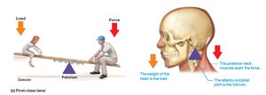
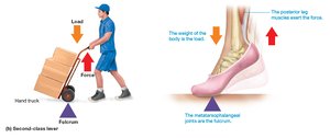
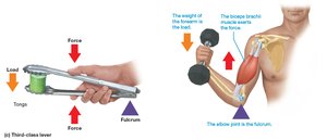
Muscles act on bones as levers, which consist of a load, applied force, and fulcrum (pivot point). There are three classes of levers:
First-class lever: Fulcrum between load and applied force; load moves in opposite direction to force.
Second-class lever: Fulcrum at one end, applied force at the other, load in between; load moves in same direction as force.
Third-class lever: Fulcrum and applied force close together at one end, load at the other; load moves in same direction as force.
Changes Caused by Physical Training
Myoplasticity refers to changes in muscle structure as a result of physical training. Most mature skeletal muscle fibers are amitotic, but satellite cells retain mitotic ability and can help repair injured muscle. Training-induced changes occur within muscle fibers, not in their number.
Endurance training: Increases frequency of motor unit activation and moderate force production. Biochemical changes include increased oxidative enzymes, mitochondria, fatigue resistance, and blood vessel network. Most dramatic in type I fibers.
Resistance (strength) training: Moderate increase in motor unit activation and large increase in force production. Anatomical changes include increased number of myofibrils and fiber diameter (hypertrophy). May decrease endurance if not balanced.
Disuse: Leads to decrease in myofibrils, fiber size, and oxidative enzymes (atrophy), resulting in reduced strength and endurance.
Equation for muscle force:
Equation for lever mechanical advantage:
Additional info: The notes have been expanded to include definitions, examples, and equations for clarity and completeness.
