 뒤로
The Anatomy and Physiology of the Eye: Structure, Function, and Visual Processing
스터디 가이드 - 스마트 노트
 자료에 맞춘 맞춤형 노트, 핵심 정의, 예시, 맥락을 확장해 제공합니다.
The Eye: Structure and Function
Accessory Structures of the Eye
The accessory structures of the eye provide protection, lubrication, and support, ensuring optimal function of the visual system. These include the eyelids, superficial epithelium, tear-producing structures, and extrinsic eye muscles.
Eyelids (Palpebrae): Continuations of the skin that blink to lubricate and protect the eye, removing dust and debris.
Eyelashes: Strong hairs that prevent foreign matter from reaching the eye's surface.
Exocrine Glands: Sebaceous glands (including tarsal glands) secrete substances to prevent eyelid adhesion; lacrimal caruncle produces thick secretions.
Conjunctiva: A thin, transparent mucous membrane covering the inner eyelids and white of the eye, highly sensitive and prone to conjunctivitis (pinkeye).
Lacrimal Apparatus: Produces, distributes, and removes tears, which contain lysozyme for antibacterial action and provide nutrients to the conjunctiva.
Extrinsic Eye Muscles
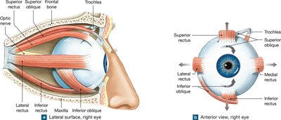
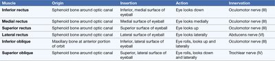
Six extrinsic eye muscles control the movement and positioning of the eyeball. These muscles originate from the orbit and insert onto the sclera.
Inferior Rectus: Moves eye downward.
Medial Rectus: Moves eye medially.
Superior Rectus: Moves eye upward.
Lateral Rectus: Moves eye laterally.
Inferior Oblique: Rolls eye upward and laterally.
Superior Oblique: Rolls eye downward and laterally.
Muscle | Origin | Insertion | Action | Innervation |
|---|---|---|---|---|
Inferior rectus | Sphenoid bone around optic canal | Inferior, medial surface of eyeball | Eye looks down | Oculomotor nerve (III) |
Medial rectus | Sphenoid bone around optic canal | Medial surface of eyeball | Eye looks medially | Oculomotor nerve (III) |
Superior rectus | Sphenoid bone around optic canal | Superior surface of eyeball | Eye looks up | Oculomotor nerve (III) |
Lateral rectus | Sphenoid bone around optic canal | Lateral surface of eyeball | Eye looks laterally | Abducens nerve (VI) |
Inferior oblique | Maxilla at anterior portion of orbit | Inferior, lateral surface of eyeball | Eye rolls, looks up and laterally | Oculomotor nerve (III) |
Superior oblique | Sphenoid bone around optic canal | Superior, lateral surface of eyeball | Eye rolls, looks down and laterally | Trochlear nerve (IV) |
Internal Anatomy of the Eye
General Structure
The eyeball is a hollow, fluid-filled organ divided into anterior and posterior cavities. The anterior cavity contains aqueous humor, while the posterior cavity (vitreous chamber) contains the vitreous body. The wall of the eye consists of three layers: fibrous, vascular, and inner (retina).
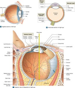
Fibrous Layer: Outermost layer, includes the sclera (white of the eye) and cornea (transparent, allows light entry).
Vascular Layer: Middle layer, includes the iris (controls pupil size), ciliary body (controls lens shape), and choroid (supplies nutrients).
Inner Layer (Retina): Innermost layer, contains photoreceptors and neurons for visual processing.
Fibrous Layer
The fibrous layer provides mechanical support, protection, and serves as an attachment site for extrinsic muscles. The sclera is dense and fibrous, while the cornea is transparent and avascular, allowing light to pass through.
Vascular Layer
The vascular layer regulates light entry, secretes aqueous humor, and controls lens shape. The iris contains smooth muscle fibers that adjust pupil diameter. Eye color is determined by the number and distribution of melanocytes and melanin granules.
Inner Layer (Retina)
The retina consists of a pigmented layer (absorbs excess light) and a neural layer (contains photoreceptors, bipolar cells, ganglion cells, and supporting cells). The neural layer processes and integrates visual information before transmitting it to the brain.
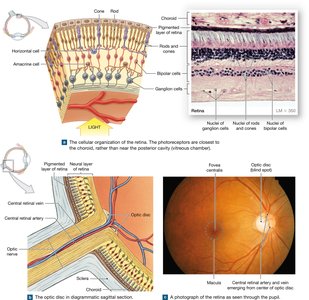
Rods: Sensitive to dim light, provide black-and-white vision.
Cones: Responsible for color vision (red, green, blue pigments), provide sharp, clear images in bright light.
ipRGCs: Non-image-forming cells influencing circadian rhythm.
Macula and Fovea Centralis: Area of highest cone concentration and sharpest vision.
Optic Disc and Blind Spot
The optic disc is the origin of the optic nerve and contains no photoreceptors, resulting in a blind spot. Involuntary eye movements and brain processing compensate for this gap in vision.
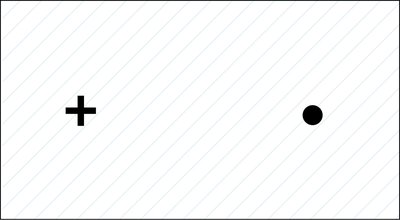
Chambers and Fluids of the Eye
Aqueous Humor
Aqueous humor is secreted by the ciliary body, circulates through the anterior and posterior chambers, and is reabsorbed via the scleral venous sinus. It maintains intraocular pressure, transports nutrients, and removes wastes.
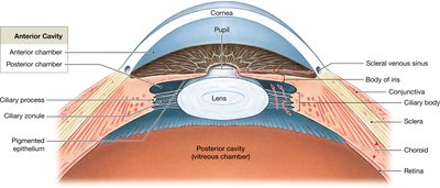
Glaucoma: Increased intraocular pressure due to impaired aqueous humor drainage, can lead to blindness.
Vitreous Body
The vitreous body is a gelatinous substance in the posterior cavity, helping maintain eye shape and holding the retina against the choroid.
The Lens and Visual Accommodation
Structure and Function of the Lens
The lens is a transparent, elastic structure held by the ciliary zonule. Its primary function is to focus light onto the retina by changing shape (accommodation).
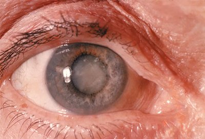
Cataracts: Loss of lens transparency due to structural changes, leading to vision impairment. Treatment involves surgical replacement.
Light Refraction and Accommodation
Light is refracted as it passes through the cornea and lens. The lens changes shape to adjust focal distance, ensuring the image is focused on the retina. Accommodation involves the lens becoming rounder for near objects and flatter for distant objects.
Focal Distance: Determined by object distance and lens shape.
Key Equations:
The focal distance () of a lens is given by:
where is the object distance and is the image distance.
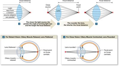
Image Formation and Visual Processing
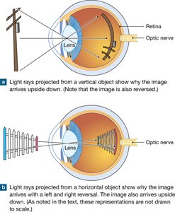
Image Reversal
The image formed on the retina is upside down and reversed left-to-right. The brain compensates for this reversal, allowing accurate perception of the environment.
Refractive Problems
Improper refraction of light leads to visual distortions such as astigmatism, myopia (nearsightedness), and hyperopia (farsightedness). These can be corrected with lenses or surgical procedures.
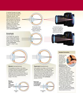
Example: In myopia, the focal point falls in front of the retina; in hyperopia, it falls behind. Astigmatism results from irregular curvature of the cornea or lens.
Additional info: The notes above expand on the original content by providing definitions, clinical context, and relevant equations for focal distance and accommodation.
