 뒤로
The Appendicular Skeleton: Structure and Principal Markings
스터디 가이드 - 스마트 노트
 자료에 맞춘 맞춤형 노트, 핵심 정의, 예시, 맥락을 확장해 제공합니다.
The Appendicular Skeleton
Overview of the Appendicular Skeleton
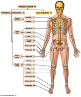
The appendicular skeleton consists of the bones that form the limbs and girdles, connecting them to the axial skeleton. It is essential for movement and manipulation of the environment, and includes the pectoral (shoulder) and pelvic (hip) girdles, as well as the bones of the upper and lower limbs.
Pectoral Girdle: Attaches the upper limbs to the trunk.
Pelvic Girdle: Attaches the lower limbs to the trunk.
Upper Limbs: Includes arm, forearm, and hand bones.
Lower Limbs: Includes thigh, leg, and foot bones.
Pectoral Girdle
Structure of the Pectoral Girdle
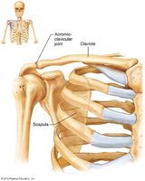
The pectoral girdle consists of two pairs of bones: the clavicles and scapulae. These bones form an almost complete circle around the upper trunk, providing attachment points for muscles and facilitating a wide range of motion for the upper limbs.
Clavicle: S-shaped bone, articulates medially with the sternum and laterally with the scapula.
Scapula: Thin, triangular bone located dorsally between ribs 2 and 7.
Glenoid Cavity: Shallow socket for the shoulder joint, allowing mobility but less stability.
Clavicle
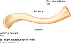
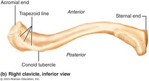
The clavicle, or collarbone, acts as a brace to keep the arms away from the body and serves as an attachment point for muscles. Its curvature ensures that fractures occur outward, away from vital structures.
Medial (sternal) end: Articulates with the sternum.
Lateral (acromial) end: Articulates with the scapula.
Principal markings: Trapezoid line, conoid tubercle.
Scapula
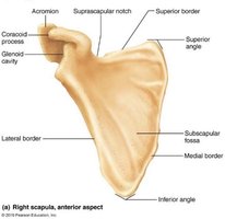
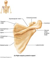
The scapula, or shoulder blade, is an irregular bone with several important features for muscle attachment and articulation. It has three borders (superior, medial, lateral) and several fossae.
Acromion: Articulates with the clavicle.
Coracoid process: Attachment for biceps muscle.
Glenoid cavity: Articulates with the head of the humerus.
Fossae: Subscapular, supraspinous, infraspinous.
Bones of the Upper Limb
Regions and Principal Markings
The upper limb is divided into three major regions: arm, forearm, and hand. Each region contains specific bones with distinct markings that serve as sites for muscle attachment and articulation.
Arm: Humerus
Forearm: Radius and ulna
Hand: Carpals, metacarpals, phalanges
Humerus
The humerus is the longest and largest bone of the upper limb, articulating with the scapula proximally and the radius and ulna distally.
Head: Fits into the glenoid cavity.
Anatomical neck vs surgical neck: Anatomical neck is just below the head; surgical neck is a common fracture site.
Tubercles: Greater and lesser, separated by the intertubercular sulcus.
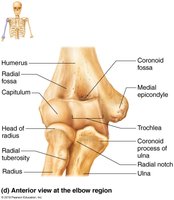
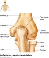
Condyles: Trochlea (articulates with ulna), capitulum (articulates with radius).
Epicondyles: Medial and lateral, sites for muscle attachment.
Radius and Ulna
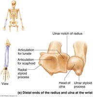
The forearm consists of two parallel bones: the radius and ulna. They articulate with each other at the proximal and distal radio-ulnar joints and are connected by the interosseous membrane.
Ulna: Forms the elbow joint, has olecranon and coronoid processes.
Radius: Contributes to the wrist joint, has a head at the proximal end and a styloid process at the distal end.
Hand
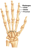
The hand consists of the carpus (wrist), metacarpus (palm), and phalanges (fingers). The carpus contains eight carpal bones arranged in two rows, while the metacarpals and phalanges form the palm and fingers, respectively.
Carpal bones: Proximal row (scaphoid, lunate, triquetrum, pisiform), distal row (trapezium, trapezoid, capitate, hamate).
Metacarpals: Five long bones, numbered I-V from thumb to little finger.
Phalanges: Four fingers have three phalanges each; the thumb (pollex) has two.
Pelvic Girdle
Structure of the Pelvic Girdle
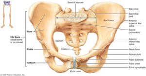
The pelvic girdle consists of two hip bones (coxal bones), which unite anteriorly and with the sacrum posteriorly. It transmits the weight of the upper body to the lower limbs and supports pelvic organs.
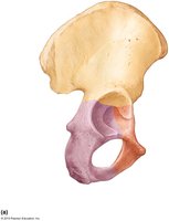
Os coxa: Each hip bone is formed by the fusion of three bones: ilium, ischium, and pubis.
Acetabulum: The socket where all three bones join, forming the hip joint.
Ilium, Ischium, and Pubis
The ilium forms the superior part of the hip bone, the ischium forms the posteroinferior part, and the pubis forms the anterior part. Each has distinct markings and features for muscle attachment and articulation.
Ilium: Iliac crest, iliac spines, pelvic brim.
Ischium: Ischial tuberosity, body, ramus.
Pubis: Pubic crest, obturator foramen, pubic symphysis.
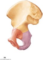
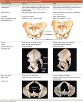
False vs True Pelvis; Male vs Female Pelves
The pelvis is divided into the false (greater) and true (lesser) pelvis. The structural differences between male and female pelves are related to childbirth and body structure.
False pelvis: Superior to the pelvic brim, supports abdominal organs.
True pelvis: Inferior to the pelvic brim, contains pelvic organs.
Male pelvis: Narrower, deeper, more robust.
Female pelvis: Wider, shallower, adapted for childbirth.
Bones of the Lower Limb
Regions and Principal Markings
The lower limb is divided into three segments: thigh, leg, and foot. It carries the entire weight of the body and is subjected to significant forces during movement.
Thigh: Femur
Leg: Tibia and fibula
Foot: Tarsals, metatarsals, phalanges
Femur
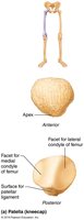
The femur is the largest, strongest, and longest bone in the body. It articulates proximally with the acetabulum of the hip and distally with the tibia and patella.
Head: Contains the fovea capitis.
Neck: Angles laterally, common site of fractures.
Trochanters: Greater and lesser, sites for muscle attachment.
Condyles and epicondyles: Articulate with tibia, sites for muscle attachment.
Patellar surface: Articulates with the patella.
Tibia and Fibula
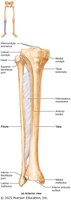
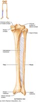
The leg contains two parallel bones: the tibia and fibula. The tibia is the main weight-bearing bone, while the fibula provides lateral stability.
Tibia: Medial and lateral condyles, tibial tuberosity, medial malleolus.
Fibula: Head, lateral malleolus, not weight-bearing.
Interosseous membrane: Connects the tibia and fibula.
Foot
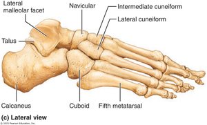
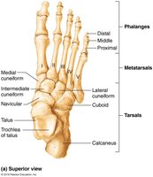
The foot consists of the tarsus (ankle), metatarsus, and phalanges (toes). The tarsus contains seven tarsal bones, with the calcaneus (heel bone) and talus being the largest and most important for weight-bearing.
Tarsal bones: Calcaneus, talus, cuboid, navicular, medial/intermediate/lateral cuneiforms.
Metatarsals: Five miniature long bones, numbered I-V from hallux to little toe.
Phalanges: Four toes have three phalanges each; the hallux has two.
Summary Table: Major Bones of the Appendicular Skeleton
Region | Main Bones | Principal Markings |
|---|---|---|
Pectoral Girdle | Clavicle, Scapula | Acromion, Glenoid cavity, Coracoid process |
Upper Limb | Humerus, Radius, Ulna, Carpals, Metacarpals, Phalanges | Head, Neck, Tuberosities, Condyles, Epicondyles |
Pelvic Girdle | Ilium, Ischium, Pubis | Iliac crest, Ischial tuberosity, Pubic symphysis |
Lower Limb | Femur, Tibia, Fibula, Tarsals, Metatarsals, Phalanges | Head, Neck, Trochanters, Condyles, Malleoli |
