 뒤로
The Axial Skeleton: Skull, Sutures, and Associated Structures
스터디 가이드 - 스마트 노트
 자료에 맞춘 맞춤형 노트, 핵심 정의, 예시, 맥락을 확장해 제공합니다.
The Integumentary System: Review of Skin Structure and Function
Layers of the Epidermis and Dermis
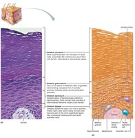
The skin is composed of two main layers: the epidermis (outer layer) and the dermis (inner layer). The epidermis consists of several sublayers, each with distinct characteristics and functions.
Stratum corneum: Outermost layer; consists of dead, keratinized cells that provide a protective barrier.
Stratum lucidum: Thin, clear layer found only in thick skin (e.g., palms, soles).
Stratum granulosum: Cells contain granules important for waterproofing and keratin formation.
Stratum spinosum: Several layers of keratinocytes; provides strength and flexibility.
Stratum basale: Deepest layer; site of cell division and source of new keratinocytes.
Dermis: Connective tissue layer containing blood vessels, nerves, and accessory structures.
Key Functions of the Skin:
Protection: Acts as a barrier against chemical, physical, and biological threats.
Body temperature regulation: Sweat glands and blood vessel constriction/dilation help maintain temperature homeostasis.
Cutaneous sensations: Contains receptors for touch, pressure, pain, and temperature.
Metabolic functions: Synthesizes vitamin D in the epidermis.
Blood reservoir: Holds approximately 5% of the body's blood volume.
Excretion: Removes waste products via sweat and sebaceous glands.
The Skeletal System: Overview and Classification
Axial and Appendicular Skeleton
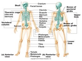
The human skeleton consists of 206 bones, accounting for about 20% of total body weight. It is divided into two main divisions:
Axial skeleton: Includes the skull, vertebral column, and rib cage. Provides central support and protects vital organs.
Appendicular skeleton: Comprises the bones of the upper and lower limbs, as well as the pectoral and pelvic girdles, which attach the limbs to the axial skeleton.
The Skull: Structure and Function
Cranial and Facial Bones
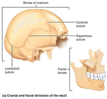
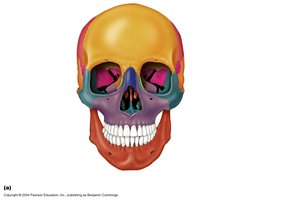
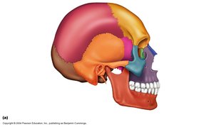
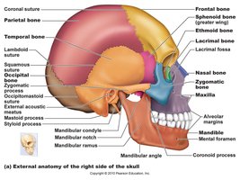
The skull is composed of 22 bones: 8 cranial bones and 14 facial bones. The cranial bones protect the brain and provide attachment sites for head and neck muscles, while the facial bones form the framework of the face and house special sense organs.
Cranial bones (8): Frontal, parietal (2), occipital, temporal (2), sphenoid, ethmoid.
Facial bones (14): Mandible, vomer, maxillae (2), zygomatic (2), nasal (2), lacrimal (2), palatine (2), inferior nasal conchae (2).
Cranial Vault and Cranial Base
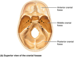
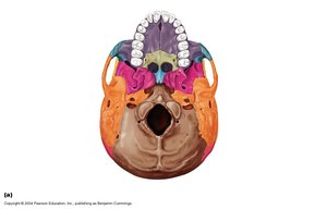
The cranial vault (calvaria) forms the superior, lateral, and posterior aspects of the skull, including the forehead. The cranial base forms the inferior aspect and is divided into three fossae: anterior, middle, and posterior. The brain sits within these fossae, enclosed by the cranial vault.
Major Cavities and Openings of the Skull
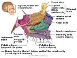
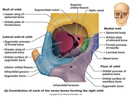
The skull contains several major cavities, including the cranial cavity, orbits, nasal cavity, and paranasal sinuses. There are 85 named openings (foramina, canals, fissures) that allow passage of nerves, blood vessels, and the spinal cord.
Key Cranial Bones
Frontal bone: Forms the forehead and superior part of the orbits.
Parietal bones: Paired bones forming the superior and lateral aspects of the skull.
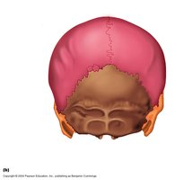
Occipital bone: Forms the posterior base of the skull.
Temporal bones: Paired bones forming the inferior and lateral aspects of the skull; house the middle and inner ear structures.
Sphenoid bone: Complex, bat-shaped bone that articulates with all other cranial bones.
Ethmoid bone: Cube-shaped bone located between the orbits and nasal cavity; features the crista galli for attachment of the brain's dura mater.
Temporal Bone: Structure and Function
The temporal bone is irregularly shaped and contains several important regions, including the squamous, tympanic, mastoid, and petrous parts. The petrous part houses the middle and inner ear, which are essential for hearing and balance.
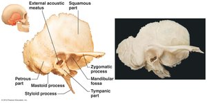
Sphenoid and Ethmoid Bones
The sphenoid bone is centrally located and articulates with all other cranial bones. The ethmoid bone is situated between the orbits and forms part of the nasal cavity.
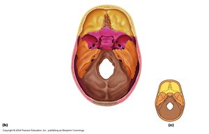
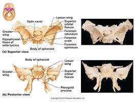
Major Cranial Sutures
Sutures are immovable joints that unite the bones of the skull. The four main sutures are:
Coronal suture: Between the frontal and parietal bones.
Squamous suture: Between the parietal and temporal bones.
Lambdoid suture: Between the occipital and parietal bones.
Sagittal suture: Between the two parietal bones.
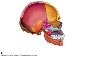
Facial Bones and Special Structures
Facial Bones
The facial bones form the structure of the face, house the special sense organs, and provide openings for air and food passage. The mandible and vomer are unpaired; all others are paired.
The Mandible
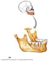
The mandible is the largest and strongest bone of the face. It forms the lower jaw and is the only movable bone of the skull (excluding the ossicles of the middle ear). Key features include the coronoid process (muscle attachment for chewing) and the mental foramen (passage for nerves and blood vessels).
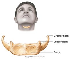
The Hyoid Bone
The hyoid bone is unique in that it does not articulate directly with any other bone. It is anchored by ligaments and serves as a movable base for the tongue and an attachment site for muscles involved in swallowing and speech. It is horseshoe-shaped and can be fractured during strangulation.
Paranasal Sinuses
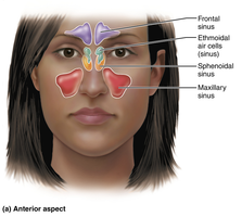
Paranasal sinuses are air-filled spaces within certain cranial bones. They lighten the skull, help warm and moisten incoming air, and contribute to voice resonance. The main sinuses are the frontal, ethmoidal, sphenoidal, and maxillary sinuses.
Summary Table: Key Skull Facts
Feature | Number |
|---|---|
Total bones in the body | 206 |
Bones in the skull | 22 |
Facial bones | 14 |
Named openings in the skull | 85 |
Main cranial sutures | 4 |
Hyoid bone | 1 |
Practice Questions
The number of bones in the human body is: 206
The number of openings in the skull is: 85
Which statement about the skull is false? Ethmoid bone is said to be bat shaped (False; the sphenoid bone is bat-shaped)
Strangulation may be expected to result in fracturing of the: Hyoid bone
Three main components of connective tissue: Ground substance, fibers, and cells
Which is found in bone but not cartilage? Blood vessels
Which glands secrete directly into the blood? Endocrine glands
Vitamin D is made from which compound on the skin? Cholesterol
Cleavage lines are: Due to collagen fibers in parallel in the dermal layer
Key Bones to Know from This Lecture
Frontal
Occipital
Temporal
Parietal
Sphenoid
Ethmoid
Mandible
Hyoid Bone
Additional info: Students should understand the general structure and function of the major cranial and facial bones, as well as the significance of sutures and the hyoid bone. Detailed knowledge of all facial bones is not required beyond the mandible for this lecture.
