 뒤로
The Axial Skeleton: Structure and Function
스터디 가이드 - 스마트 노트
 자료에 맞춘 맞춤형 노트, 핵심 정의, 예시, 맥락을 확장해 제공합니다.
The Axial Skeleton
Overview of the Axial Skeleton
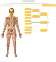
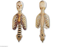
The axial skeleton forms the longitudinal axis of the human body and consists of 80 bones. It provides support and protection for the brain, spinal cord, and organs in the thorax. The axial skeleton includes the skull, vertebral column, and thoracic cage.
Skull: Composed of 8 cranial bones and 14 facial bones, plus 6 auditory ossicles and the hyoid bone.
Vertebral Column: Contains 24 vertebrae, the sacrum, and the coccyx.
Thoracic Cage: Includes 24 ribs and the sternum.
The Skull
Cranial and Facial Bones
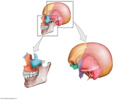
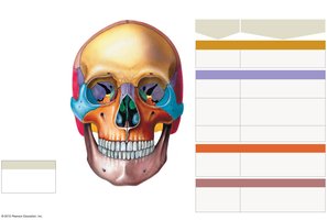
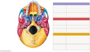
The skull is divided into cranial bones, which protect the brain, and facial bones, which support the entrances to the digestive and respiratory tracts.
Cranial Bones: Frontal, parietal, temporal, occipital, sphenoid, and ethmoid bones.
Facial Bones: Maxillae, palatine, nasal, vomer, inferior nasal conchae, zygomatic, lacrimal, and mandible.
Auditory Ossicles and Hyoid Bone
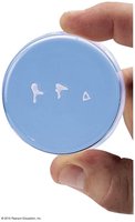
The auditory ossicles are three tiny bones in the middle ear (malleus, incus, stapes) that transfer sound from the tympanic membrane to the inner ear. The hyoid bone supports the tongue and is not directly attached to other bones of the skull.
Major Sutures of the Skull
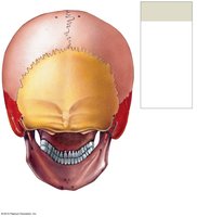
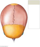
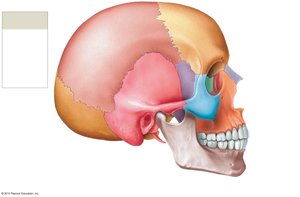
Sutures are immovable joints that connect the bones of the skull. The four major sutures are:
Lambdoid Suture: Separates the occipital from parietal bones.
Coronal Suture: Attaches the frontal bone to the parietal bones.
Sagittal Suture: Between the parietal bones, from lambdoid to coronal suture.
Squamous Suture: Forms boundaries between temporal and parietal bones.
Key Foramina of the Skull
Foramina are openings in the skull that allow the passage of nerves and blood vessels. Examples include:
Supraorbital foramen: Passage for the supraorbital nerve and artery.
Optic canal: Passage for the optic nerve (CN II) and ophthalmic artery.
Foramen magnum: Passage for the medulla oblongata, vertebral arteries, and accessory nerve (CN XI).
Cranial Bones: Structure and Landmarks
Occipital and Parietal Bones
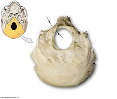
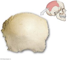
The occipital bone forms the posterior and base of the skull, containing the foramen magnum and occipital condyles. The parietal bones form the superior and lateral aspects of the cranium.
Frontal Bone
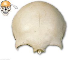
The frontal bone forms the forehead and the superior part of the orbit. Key features include the frontal squama, supraorbital margin, lacrimal fossa, and frontal sinuses.
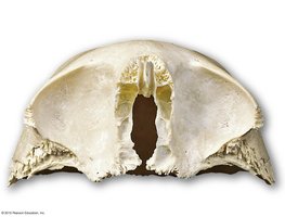
Temporal Bones
The temporal bones form the sides and base of the cranium. They contain the external acoustic meatus, mastoid process, styloid process, and zygomatic process.
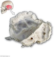
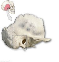
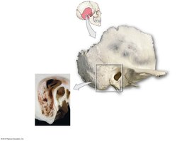
Sphenoid and Ethmoid Bones
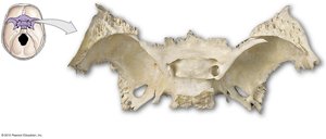
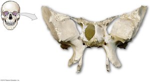
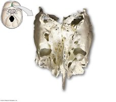
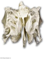
The sphenoid bone is a complex bone at the base of the skull, containing the sella turcica and several foramina. The ethmoid bone forms part of the nasal cavity and the medial wall of the orbit, containing the cribriform plate and crista galli.
Facial Bones: Structure and Landmarks
Maxillae and Palatine Bones
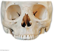
The maxillae form the upper jaw and most of the hard palate. The palatine bones form the posterior part of the hard palate and part of the nasal cavity and orbit.
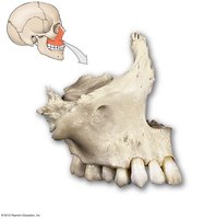
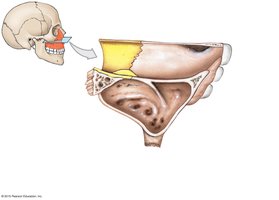
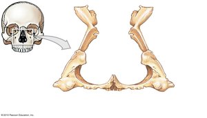
Nasal, Lacrimal, and Zygomatic Bones
The nasal bones form the bridge of the nose. The lacrimal bones are small bones in the medial wall of each orbit. The zygomatic bones form the cheeks and part of the orbit.
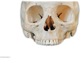
Mandible
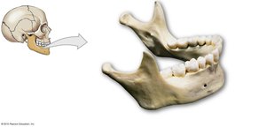
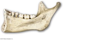
The mandible is the lower jawbone and the only movable bone of the skull. It contains the mental foramen, mandibular foramen, and alveolar processes for teeth attachment.
Hyoid Bone
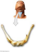
The hyoid bone is located in the neck between the mandible and larynx. It supports the tongue and serves as an attachment for muscles associated with swallowing.
The Vertebral Column
Structure and Function
The vertebral column, or spine, protects the spinal cord and supports the head and body. It consists of 26 bones: 24 vertebrae, the sacrum, and the coccyx. The vertebral column is divided into regions based on anatomical characteristics and function.
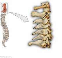
Cervical (C1–C7): Neck region, supports the head.
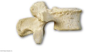
Thoracic (T1–T12): Upper back, articulates with ribs.
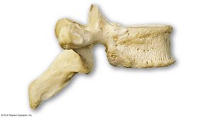
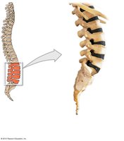
Lumbar (L1–L5): Lower back, supports the weight of the trunk.
Sacral and Coccygeal: Sacrum and coccyx at the base of the spine.
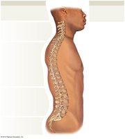
Vertebral Anatomy
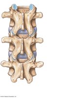
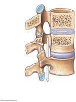
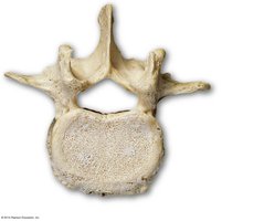
A typical vertebra consists of a vertebral body, vertebral arch (pedicles and laminae), and articular processes. The vertebral foramen forms the vertebral canal, which encloses the spinal cord. Intervertebral discs of fibrocartilage separate vertebral bodies and absorb shock.
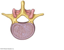
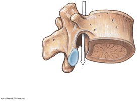
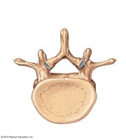
Regional Differences in Vertebrae
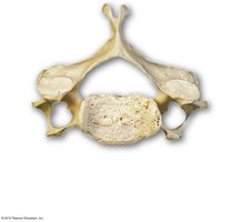
Cervical Vertebrae: Small body, large vertebral foramen, bifid spinous process (except C1), transverse foramina.
Thoracic Vertebrae: Heart-shaped body, costal facets for rib articulation, long spinous process.
Lumbar Vertebrae: Largest bodies, short and thick spinous processes, no costal facets.
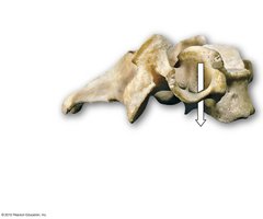
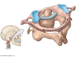
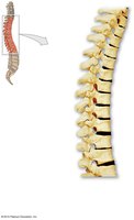
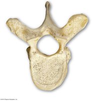
The Sacrum and Coccyx
Structure and Function
The sacrum is a large, triangular bone at the base of the spine, formed by the fusion of five sacral vertebrae. It articulates with the fifth lumbar vertebra and the coccyx. The coccyx, or tailbone, is composed of three to five fused vertebrae.
The Thoracic Cage
Structure and Function
The thoracic cage, or rib cage, supports the thoracic cavity and protects the heart and lungs. It consists of the thoracic vertebrae, ribs, and sternum.
True Ribs (1–7): Attach directly to the sternum via costal cartilages.
False Ribs (8–12): Do not attach directly to the sternum. Ribs 8–10 are vertebrochondral, and ribs 11–12 are floating ribs.
Structures of the Ribs
Each rib has a head (capitulum) with articular facets, a neck, a tubercle with a facet for articulation with the transverse process of a vertebra, and a shaft (body) that attaches to muscles.
The Sternum
The sternum is a flat bone in the midline of the thoracic wall, consisting of three parts:
Manubrium: Superior portion, articulates with clavicles and first rib pair, contains the jugular notch.
Sternal Body: Attaches to the manubrium and costal cartilages of ribs 2–7.
Xiphoid Process: Smallest part, attaches to the sternal body, diaphragm, and rectus abdominis muscles.
Additional info: The axial skeleton is essential for protecting vital organs, supporting the body, and providing attachment points for muscles involved in movement and respiration.
