 뒤로
The Axial Skeleton: Structure, Classification, and Key Features
스터디 가이드 - 스마트 노트
 자료에 맞춘 맞춤형 노트, 핵심 정의, 예시, 맥락을 확장해 제공합니다.
The Skeletal System: Divisions and Overview
Divisions of the Skeletal System
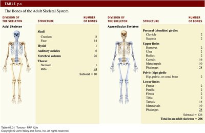
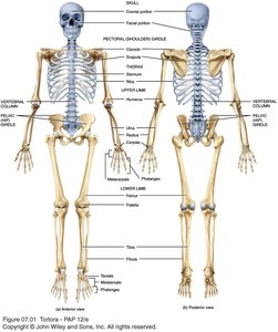
The human skeleton is composed of 206 named bones, grouped into two principal divisions: the axial skeleton and the appendicular skeleton. The axial skeleton consists of bones that lie around the longitudinal axis of the body, including the skull, auditory ossicles, hyoid bone, ribs, sternum, and vertebral column. The appendicular skeleton comprises the bones of the upper and lower limbs, as well as the girdles that connect them to the axial skeleton.
Axial Skeleton: Provides support and protection for the brain, spinal cord, and organs in the thoracic cavity.
Appendicular Skeleton: Facilitates movement and interaction with the environment.
Types of Bones
Classification by Shape
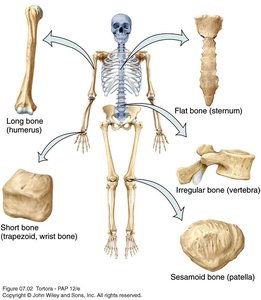
Bones are classified into five main types based on their shape, each serving distinct structural and functional roles.
Long Bones: Greater length than width; slightly curved for strength. Examples: femur, humerus, phalanges.
Short Bones: Cube-shaped, nearly equal in length and width. Examples: carpals, tarsals.
Flat Bones: Thin, composed of two parallel plates of compact bone enclosing spongy bone. Examples: cranial bones, sternum, ribs.
Irregular Bones: Complex shapes that do not fit other categories. Examples: vertebrae, hip bones, some facial bones.
Sesamoid Bones: Small, round bones embedded in tendons; protect tendons from wear. Example: patella.
Sutural Bones: Small bones located in sutures of cranial bones (not always counted in the total bone number).
Bone Surface Markings
Depressions, Openings, and Processes
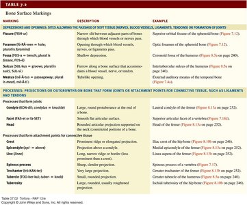
Bones display characteristic surface markings, which are structural features adapted for specific functions. These markings are classified as:
Depressions and Openings: Allow passage of blood vessels and nerves or form joints. Examples include foramina, fissures, sulci, and fossae.
Processes: Projections or outgrowths that form joints or serve as attachment points for ligaments and tendons. Examples include condyles, heads, trochanters, tubercles, and spines.
Skull: Structure and Function
Cranial and Facial Bones
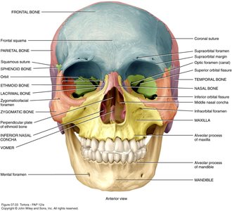
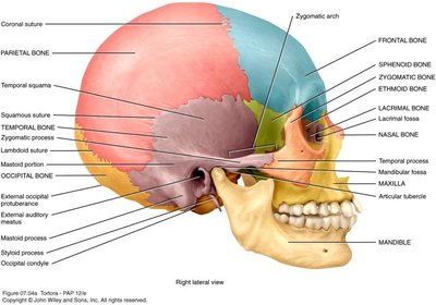
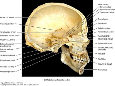
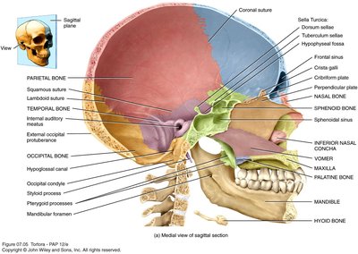
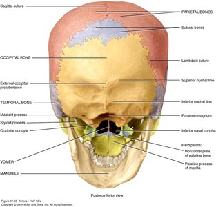
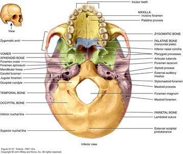
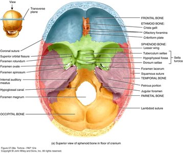
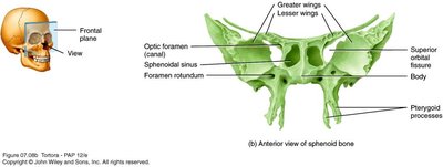
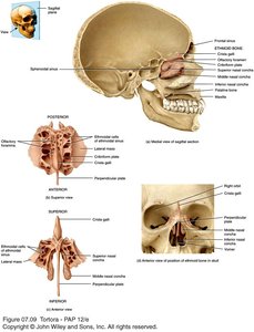
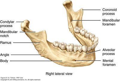
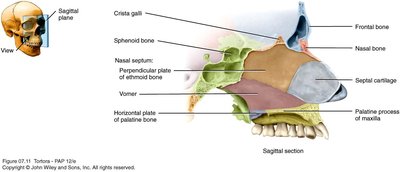
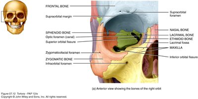
The skull consists of 22 bones, grouped into cranial and facial categories. Cranial bones form the cranial cavity and protect the brain, while facial bones form the structure of the face and support entrances to the digestive and respiratory systems.
Cranial Bones (8): Frontal, parietal (2), temporal (2), occipital, sphenoid, ethmoid.
Facial Bones (14): Nasal (2), maxillae (2), zygomatic (2), mandible, lacrimal (2), palatine (2), inferior nasal conchae (2), vomer.
Functions of the Skull
Protects and supports special sense organs and the brain.
Forms cavities for the nasal cavity, orbits, paranasal sinuses, and small cavities for hearing and equilibrium.
Provides attachment points for muscles involved in head movement and facial expression.
Facial bones support entrances to the digestive and respiratory systems.
Unique Features of the Skull
Sutures: Immovable joints that fuse most skull bones together.
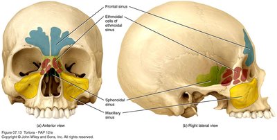
Paranasal Sinuses: Cavities within cranial and facial bones near the nasal cavity; serve as resonating chambers for sound.
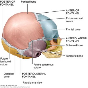
Fontanels: Areas of unossified tissue in the fetal skull, providing flexibility during birth; eventually replaced by bone.
Hyoid Bone
Structure and Function
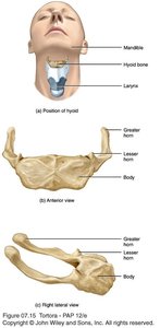
The hyoid bone is unique because it does not articulate with any other bone. It supports the tongue and provides attachment sites for muscles of the tongue, neck, and pharynx. It also helps keep the larynx open.
Location: Situated in the anterior neck, between the mandible and larynx.
Function: Assists in swallowing and speech.
Vertebral Column
Structure and Functions
The vertebral column, also known as the spine or backbone, is composed of 26 vertebrae in adults. It protects the spinal cord, supports the head, and serves as a point of attachment for the ribs, pelvic girdle, and muscles.
Curves: Increase strength, maintain balance, absorb shocks, and protect vertebrae from fracture.
Regions: Cervical (7), thoracic (12), lumbar (5), sacrum (1, fused), coccyx (1, fused).
Conditions: Kyphosis, lordosis, and scoliosis are abnormal curvatures.
Intervertebral Discs
Located between adjacent vertebrae.
Form strong joints, permit movement, and absorb vertical shock.
Vertebrae consist of a body (weight-bearing), vertebral arch (surrounds spinal cord), and several processes (muscle attachment).
Thorax
Structure and Functions
The thoracic cage is formed by the sternum, ribs, costal cartilages, and thoracic vertebrae. It encloses and protects organs in the thoracic and abdominal cavities, supports the bones of the upper limbs, and plays a role in breathing.
Sternum: Breastbone, consists of manubrium, body, and xiphoid process.
Ribs: Twelve pairs provide structural support to the thoracic cavity.
Costal Cartilages: Contribute to the elasticity of the thoracic cage.
Summary Table: Bones of the Adult Skeletal System
Division of the Skeleton | Structure | Number of Bones |
|---|---|---|
Axial Skeleton | Skull (Cranium, Face), Hyoid, Auditory ossicles, Vertebral column, Thorax (Sternum, Ribs) | 80 |
Appendicular Skeleton | Pectoral girdles, Upper limbs, Pelvic girdle, Lower limbs | 126 |
Total in adult skeleton | 206 | |
Summary Table: Bone Surface Markings
Marking | Description | Example |
|---|---|---|
Foramen | Opening for blood vessels, nerves, or ligaments | Foramen magnum of occipital bone |
Fossa | Shallow depression | Coronoid fossa of humerus |
Condyle | Large, round protuberance at end of bone | Medial condyle of femur |
Process | Prominent projection | Mastoid process of temporal bone |
Spine | Sharp, slender projection | Ischial spine of hip bone |
Additional info: The notes above expand on the original content with academic context, definitions, and examples to ensure completeness and clarity for college-level Anatomy & Physiology students.
