 뒤로
The Brain: Structure, Function, and Organization
스터디 가이드 - 스마트 노트
 자료에 맞춘 맞춤형 노트, 핵심 정의, 예시, 맥락을 확장해 제공합니다.
The Major Regions of the Brain
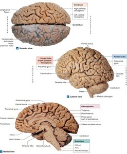
Overview of Brain Structure
The adult human brain is a highly complex organ, containing approximately 20 billion neurons organized into hundreds of neuronal pools. It is responsible for all conscious and unconscious activities, including thought, sensation, memory, and movement. The brain is divided into six major regions, each with distinct functions and anatomical features.
Cerebrum: The largest region, responsible for conscious thought, intellectual functions, memory, and voluntary movement.
Diencephalon: Contains relay and processing centers for sensory information and is involved in emotion and hormone production.
Midbrain: Processes visual and auditory information and maintains consciousness.
Pons: Connects the cerebellum to the brainstem and is involved in motor control.
Medulla Oblongata: Regulates autonomic functions such as heart rate, blood pressure, and respiration.
Cerebellum: Adjusts motor activities based on sensory information and stored memories.
The Ventricles of the Brain
Structure and Function of Ventricles
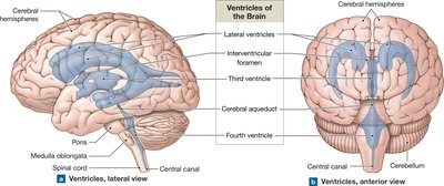
The brain contains four internal chambers called ventricles, which are filled with cerebrospinal fluid (CSF). These ventricles are essential for cushioning the brain, supporting its weight, and facilitating the transport of nutrients and waste.
Lateral Ventricles: Located in each cerebral hemisphere; communicate with the third ventricle via the interventricular foramen.
Third Ventricle: Located in the diencephalon; connects to the fourth ventricle via the cerebral aqueduct.
Fourth Ventricle: Extends into the pons and medulla oblongata; continuous with the central canal of the spinal cord.
Cerebrospinal Fluid (CSF)
Formation and Circulation
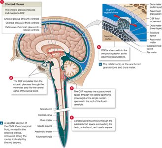
Cerebrospinal fluid is produced by the choroid plexus in each ventricle and circulates throughout the CNS. CSF serves to cushion the brain, support its weight, and transport nutrients and waste.
Production: Choroid plexus produces CSF at a rate of about 500 mL/day.
Circulation: CSF flows through the ventricles, enters the subarachnoid space, and is absorbed into the venous circulation via arachnoid granulations.
Clinical Relevance: Changes in CSF composition can indicate CNS injury or disease; lumbar puncture is used for diagnostic sampling.
The Cerebrum
Structure of the Cerebral Hemispheres
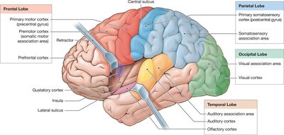
The cerebrum is covered by a layer of gray matter called the cerebral cortex, which forms folds (gyri) separated by sulci and fissures. The cortex is divided into lobes named after the overlying skull bones: frontal, parietal, temporal, and occipital.
Gyri: Increase surface area and neuron number.
Lobes: Each lobe has specialized functions related to sensory and motor processing.
Hemispheres: Left and right hemispheres control opposite sides of the body.
Motor and Sensory Areas
Primary Motor Cortex: Located in the precentral gyrus; controls voluntary movements.
Primary Somatosensory Cortex: Located in the postcentral gyrus; receives sensory information.
Visual, Auditory, Gustatory, and Olfactory Cortices: Specialized regions for processing sight, sound, taste, and smell.
Association Areas
Somatosensory Association Area: Interprets sensory information.
Premotor Cortex: Coordinates learned movements.
Visual and Auditory Association Areas: Interpret sensory data from respective cortices.
Cortical Connections
Corpus Callosum: Major bundle of axons connecting the two hemispheres.
Other White Matter Tracts: Link cortex with diencephalon, brainstem, cerebellum, and spinal cord.
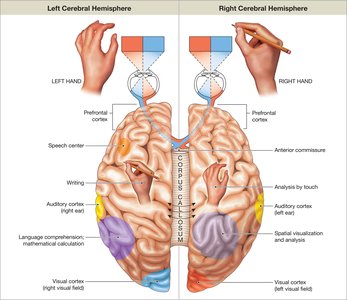
Hemispheric Lateralization
Functional Specialization
Each cerebral hemisphere is specialized for certain functions, a phenomenon known as hemispheric lateralization.
Left Hemisphere: Language skills, analytical tasks, mathematical calculations, and logical decision making.
Right Hemisphere: Spatial relationships, sensory analysis, emotional context, and recognition of faces.
Corpus Callosum: Ensures communication between hemispheres.
The Electroencephalogram (EEG)
Brain Waves and Clinical Applications
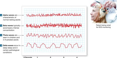
The electrical activity of the brain can be measured using an electroencephalogram (EEG). EEG records brain waves, which are correlated with levels of consciousness and can be used to diagnose brain disorders.
Alpha Waves: Normal resting adults.
Beta Waves: Accompany intense concentration.
Theta Waves: Seen in children and frustrated adults.
Delta Waves: Occur in deep sleep and certain pathological conditions.
Clinical Note: Aphasia and Dyslexia
Language Disorders
Aphasia: Disorder affecting the ability to speak or read, often resulting from damage to language areas in the cortex.
Dyslexia: Difficulty in reading and writing, often due to problems in processing visual or auditory information.

Memory
Types and Storage of Memory
Memory is classified as fact memory (specific information) and skill memory (learned motor behaviors).
Short-term Memory: Temporary, easily recalled information.
Long-term Memory: Lasts much longer, sometimes a lifetime; stored mainly in the cerebral cortex.
Memory Consolidation: Conversion from short-term to long-term memory.
Amnesia: Loss of memory due to disease or trauma.
The Basal Nuclei
Subconscious Motor Control
The basal nuclei are masses of gray matter deep within the cerebral hemispheres. They coordinate learned movement patterns and control skeletal muscle tone at the subconscious level.
Caudate Nucleus: Follows the curve of the lateral ventricle.
Lentiform Nucleus: Includes globus pallidus and putamen.
Amygdaloid Body: Part of the limbic system, involved in emotion.
The Limbic System
Emotion and Memory
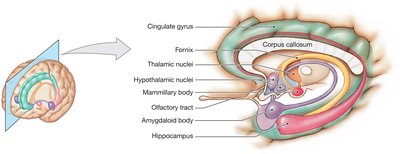
The limbic system is a functional grouping of structures involved in emotion, motivation, and long-term memory.
Amygdaloid Bodies: Link emotions with memories and regulate heart rate and fear responses.
Hippocampus: Essential for learning and memory storage.
Mammillary Bodies: Process olfactory sensations and control eating reflexes.
Fornix: Connects hippocampus with hypothalamus.
The Diencephalon
Integration and Relay Centers
The diencephalon surrounds the third ventricle and consists of the epithalamus, thalamus, and hypothalamus.
Epithalamus: Contains the pineal gland, which secretes melatonin for regulating day-night cycles.
Thalamus: Final relay point for ascending sensory information; filters and coordinates motor commands.
Hypothalamus: Controls autonomic functions, hormone production, hunger, thirst, body temperature, and daily activity cycles.
The Brainstem
Midbrain, Pons, and Medulla Oblongata
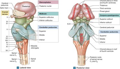
The brainstem is composed of the midbrain, pons, and medulla oblongata, each with specialized functions.
Midbrain: Processes visual and auditory information; contains the reticular formation and substantia nigra.
Pons: Links cerebellum with other brain regions; involved in respiration and motor control.
Medulla Oblongata: Connects brain with spinal cord; regulates vital autonomic functions such as heart rate and respiration.
The Cerebellum
Motor Coordination and Balance
The cerebellum is responsible for adjusting postural muscles and fine-tuning movements. It compares motor commands with proprioceptive information to ensure smooth, coordinated actions.
Cerebellar Peduncles: Tracts linking cerebellum with other brain regions.
Ataxia: Disturbance in balance due to cerebellar damage.
Summary Table: Major Brain Regions and Functions
Region | Main Function |
|---|---|
Cerebrum | Conscious thought, memory, voluntary movement |
Diencephalon | Sensory relay, emotion, hormone production |
Midbrain | Visual/auditory processing, consciousness |
Pons | Motor control, respiration |
Medulla Oblongata | Autonomic regulation, relay station |
Cerebellum | Motor coordination, balance |
Key Equations
CSF Production Rate:
CSF Volume Replacement:
Additional info:
Some details about the limbic system and basal nuclei were inferred for completeness.
Clinical notes on aphasia, dyslexia, and seizures were expanded for academic context.
