 뒤로
뒤로The Cardiovascular System: The Heart – Anatomy & Physiology Study Guide
스터디 가이드 - 스마트 노트
 자료에 맞춘 맞춤형 노트, 핵심 정의, 예시, 맥락을 확장해 제공합니다.
The Heart: Size and Location
General Overview
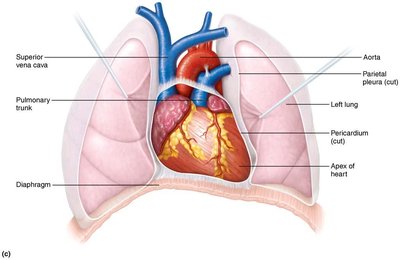
The heart is a muscular organ essential for pumping blood throughout the body. It is located in the mediastinum, positioned obliquely with its apex directed toward the left hip.
Size: 250-350 g (less than 1 lb), 12-14 cm long.
Position: Two-thirds of its mass lies to the left of the midsternal line.
The Pericardium and Heart Wall
Layers of the Heart
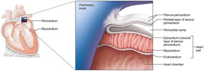
The heart is surrounded and protected by several layers, each with distinct functions.
Fibrous pericardium: Dense irregular connective tissue that protects and anchors the heart.
Parietal layer of serous pericardium: Lines the internal surface of the fibrous pericardium.
Pericardial cavity: Space containing serous fluid to reduce friction.
Epicardium: Visceral layer of the serous pericardium, forms the outermost layer of the heart wall.
Myocardium: The thick, muscular middle layer responsible for contraction.
Endocardium: The innermost endothelial lining supported by thin connective tissue.
Myocardium: Structure and Function
Cardiac Muscle Bundles
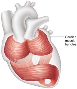
The myocardium consists of branching cardiac muscle cells connected by connective tissue fibers (collagen and elastin), arranged in circular bundles.
Support: Ropelike rings of muscle provide structural support around the base of vessels and valves.
Electrical Pathways: Embedded connective tissue guides the spread of action potentials.
Chambers of the Heart
Atria: Receiving Chambers
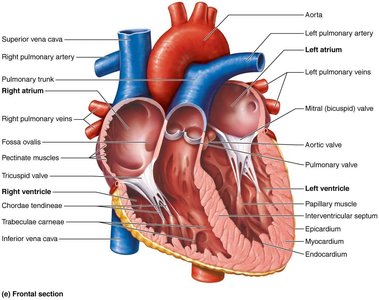
The atria are thin-walled chambers that receive blood returning to the heart.
Right Atrium: Receives blood from the superior vena cava, inferior vena cava, and coronary sinus.
Left Atrium: Receives blood from the right and left pulmonary veins.
Ventricles: Discharging Chambers
The ventricles are thick-walled chambers that pump blood out of the heart.
Right Ventricle: Pumps blood into the pulmonary trunk, which divides into right and left pulmonary arteries.
Left Ventricle: Pumps blood into the aorta.
Blood Flow Through the Heart: Two Circuits
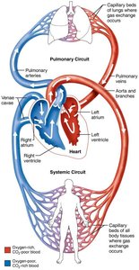
Pulmonary and Systemic Circuits
The heart operates two main circulatory circuits:
Pulmonary Circuit: Right side of the heart; pumps blood to the lungs for oxygenation. Shorter, low pressure.
Systemic Circuit: Left side of the heart; pumps oxygenated blood to the rest of the body. Higher pressure, more friction.
Heart Valves: Ensuring One-Way Blood Flow
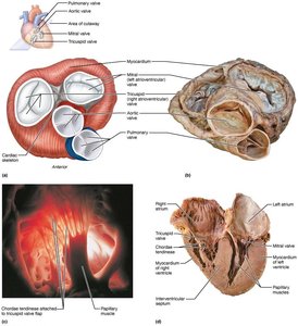
Atrioventricular (AV) Valves
AV valves prevent backflow from the ventricles into the atria.
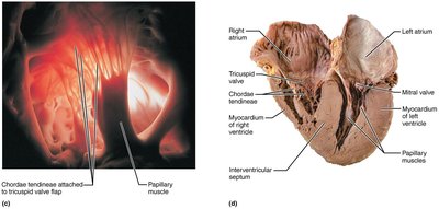
Tricuspid Valve: Right AV valve with three cusps.
Mitral (Bicuspid) Valve: Left AV valve with two cusps.
Chordae Tendineae: "Heart strings" anchor valve cusps to papillary muscles, preventing eversion during contraction.
Papillary Muscles: Contract prior to ventricular contraction, taking up slack in the chordae tendineae.
Semilunar Valves
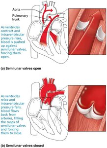
Semilunar valves prevent backflow from arteries into the ventricles.
Aortic Valve: Between left ventricle and aorta.
Pulmonary Valve: Between right ventricle and pulmonary trunk.
Each valve has three pocket-like cusps that close when filled with blood.
Microscopic Anatomy of Cardiac Muscle
Cardiac Muscle Cell Structure
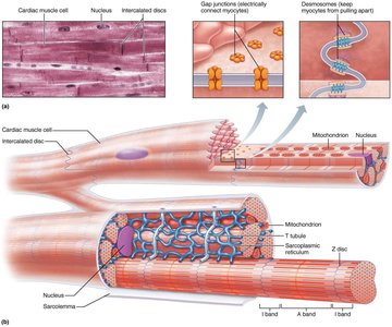
Cardiac muscle cells are short, wide, branched, striated, and interlocking, typically with one or two nuclei.
T tubules: Present, but no terminal cisternae.
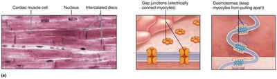
Intercalated Discs: Specialized junctions containing desmosomes and gap junctions.
Desmosomes: Attach muscle cells and prevent tearing.
Gap Junctions: Allow ions to pass, electrically coupling cells.
Functional Syncytium: The myocardium acts as a single coordinated unit.
Mitochondria: Large and abundant (25-35% of cell volume).
Skeletal vs. Cardiac Muscle Cells
Key Differences
1% of cardiac muscle cells are self-excitable (automaticity).
Heart contracts as one unit; no neuromuscular junctions or motor units.
Depolarization opens slow calcium channels, triggering further calcium release from the sarcoplasmic reticulum.
Absolute refractory period is as long as contraction (200 ms); prevents summation and tetanus.
Heart relies on aerobic respiration; anaerobic conditions lead to decreased ATP, increased H+, failed Ca pumps, and closure of gap junctions.
Pacemaker Cells and Action Potentials
Pacemaker Cell Physiology
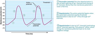
Pacemaker cells have an unstable resting membrane potential.
Slow Depolarization: Na+ entry creates gradual depolarization toward threshold.
Threshold: At -40 mV, Ca2+ rushes in, generating the action potential.
Cardiac Conduction System
SA Node and Impulse Transmission
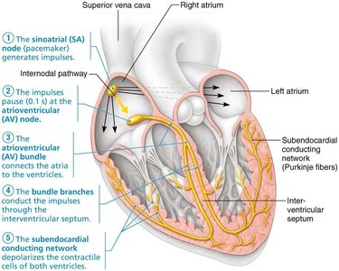
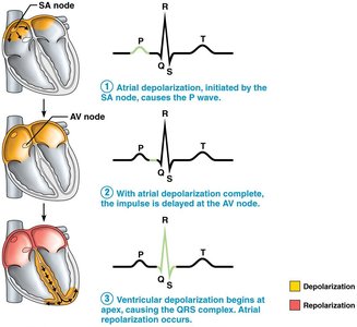
The sinoatrial (SA) node, located in the right atrial wall, sets the sinus rhythm at approximately 75 beats per minute.
Impulse is delayed 0.1 seconds between SA and AV nodes, allowing atria to contract before ventricles.
Purkinje fibers are more elaborate in the left ventricle; papillary muscles contract first, then the ventricle.
Impulse transmission from SA node to Purkinje fibers takes about 0.22 seconds.
Autonomic Innervation of the Heart
Parasympathetic and Sympathetic Regulation
The heart receives autonomic innervation from the cardiac center in the medulla oblongata.
Parasympathetic: Vagus nerve releases acetylcholine (ACh) to SA and AV nodes, slowing heart rate (vagal tone).
Sympathetic: Cardiac center stimulates T1-T5 preganglionic neurons, activating β1 receptors in SA and AV nodes and heart muscle, increasing heart rate.
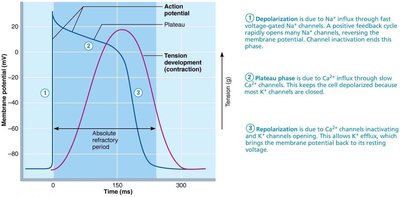
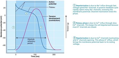
Action Potential of Contractile Cardiac Muscle Cells
Phases of Action Potential
Depolarization: Rapid Na+ influx creates the action potential.
Plateau: Ca2+ influx prolongs the action potential, ensuring sustained contraction for efficient blood ejection.
Repolarization: K+ efflux returns the membrane to resting potential.
Electrocardiography (ECG/EKG)
ECG Tracing and Heart Sounds
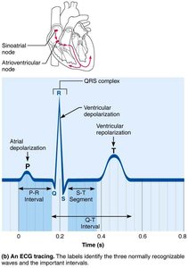
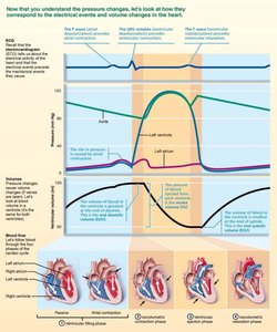
An ECG is a composite of all action potentials generated by the heart.
P wave: Atrial depolarization.
QRS complex: Ventricular depolarization.
T wave: Ventricular repolarization.
Heart Sounds: "Lub" (AV valves close), "Dup" (SL valves close).
Systole: Contraction phase.
Diastole: Relaxation phase.
The Cardiac Cycle
Phases of the Cardiac Cycle
The cardiac cycle (0.8 seconds) consists of four phases:
Ventricular Filling: Atria and ventricles relax, AV valves open, 80% of ventricular filling occurs. Atrial systole propels residual blood; end diastolic volume (EDV) is highest.
Isovolumetric Contraction: Ventricular depolarization precedes contraction; AV valves close as pressure builds.
Ventricular Ejection: When ventricular pressure exceeds aortic pressure, SL valves open and blood is ejected; stroke volume is the amount ejected.
Isovolumetric Relaxation: Ventricular relaxation, SL valves close (dicrotic notch), end systolic volume (ESV) is lowest.
Cardiac Output and Regulation
Cardiac Output (CO)
Cardiac output is the amount of blood pumped by each ventricle per minute.
Formula:
Normal CO: 75 beats/min × 70 ml/beat = 5250 ml/min (5.25 L/min)
Cardiac Reserve: Maximal CO – Resting CO
Factors Influencing Stroke Volume
Preload: Degree of cardiac muscle stretch before systole; increased preload increases stroke volume (Frank-Starling law).
Contractility: Contractile strength independent of preload; increased by epinephrine, thyroxine, and calcium.
Afterload: Back pressure exerted on the aortic/pulmonary valves; increased afterload (e.g., hypertension) reduces stroke volume.
Regulation of Heart Rate
Autonomic Regulation: Sympathetic increases HR, parasympathetic decreases HR.
Chemical Regulation: Epinephrine and thyroxine increase HR; regular exercise increases fitness, decreases HR, and increases EDV.
Summary Table: Heart Wall Layers
Layer | Location | Function |
|---|---|---|
Fibrous Pericardium | Outermost | Protects and anchors heart |
Parietal Serous Pericardium | Inner surface of fibrous pericardium | Lines fibrous pericardium |
Pericardial Cavity | Between parietal and visceral layers | Contains serous fluid |
Epicardium | Visceral layer of serous pericardium | Outer layer of heart wall |
Myocardium | Middle layer | Muscular, contracts to pump blood |
Endocardium | Innermost | Lines heart chambers |
Summary Table: Heart Valves
Valve | Location | Function |
|---|---|---|
Tricuspid (AV) | Right atrium/ventricle | Prevents backflow to atrium |
Mitral (AV) | Left atrium/ventricle | Prevents backflow to atrium |
Aortic (SL) | Left ventricle/aorta | Prevents backflow to ventricle |
Pulmonary (SL) | Right ventricle/pulmonary trunk | Prevents backflow to ventricle |
Summary Table: Cardiac Cycle Phases
Phase | Event | Key Feature |
|---|---|---|
Ventricular Filling | AV valves open, blood flows in | EDV highest |
Isovolumetric Contraction | AV valves close, pressure builds | All chambers closed |
Ventricular Ejection | SL valves open, blood ejected | Stroke volume |
Isovolumetric Relaxation | SL valves close, ventricles relax | ESV lowest |
Summary Table: Cardiac Output Factors
Factor | Effect on Stroke Volume |
|---|---|
Preload | ↑ Preload → ↑ SV |
Contractility | ↑ Contractility → ↑ SV |
Afterload | ↑ Afterload → ↓ SV |
Key Equations
Cardiac Output:
Stroke Volume:
Additional info:
Frank-Starling law states that increased venous return stretches cardiac muscle, leading to increased force of contraction and stroke volume.
Heart sounds are clinically important for diagnosing valve function.
ECG interpretation is fundamental for assessing cardiac electrical activity and diagnosing arrhythmias.
