 뒤로
뒤로The Cardiovascular System: The Heart – Structure, Function, and Physiology
스터디 가이드 - 스마트 노트
 자료에 맞춘 맞춤형 노트, 핵심 정의, 예시, 맥락을 확장해 제공합니다.
The Cardiovascular System: The Heart
Cardiac Muscle Contraction
The contraction of cardiac muscle is essential for the heart's function as a pump. Cardiac muscle cells exhibit unique properties that distinguish them from skeletal muscle cells.
Automaticity: About 1% of cardiac cells are self-excitable, meaning they can initiate their own depolarization.
Gap Junctions: These allow electrical impulses to pass rapidly between cells, ensuring the heart contracts as a unit.
Long Absolute Refractory Period: Cardiac muscle has a refractory period of about 250 ms, preventing tetanus and ensuring rhythmic contractions.
Depolarization opens voltage-gated fast Na+ channels, causing a rapid reversal of membrane potential from –90 mV to +30 mV. The depolarization wave in T tubules triggers the sarcoplasmic reticulum (SR) to release Ca2+, and slow Ca2+ channels in the sarcolemma open, prolonging the depolarization phase (plateau).
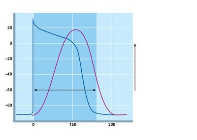
Excitation-Contraction Coupling: Ca2+ binds to troponin, initiating filament sliding and contraction. The action potential and contractile phase are longer in cardiac muscle than in skeletal muscle. Repolarization occurs when Ca2+ channels inactivate and voltage-gated K+ channels open.
Heart Physiology: Electrical Events
The heart's electrical activity is coordinated by the intrinsic cardiac conduction system, a network of autorhythmic cells that initiate and distribute impulses.
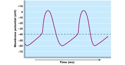
Autorhythmic Cells: These cells have unstable resting potentials (pacemaker potentials) due to open slow Na+ channels.
At threshold, voltage-gated Ca2+ channels open, causing explosive Ca2+ influx and depolarization.
Repolarization results from Ca2+ channel inactivation and K+ channel opening.
Sequence of Excitation in the Heart
The heart's electrical impulses follow a precise pathway to ensure coordinated contraction:
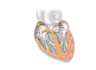
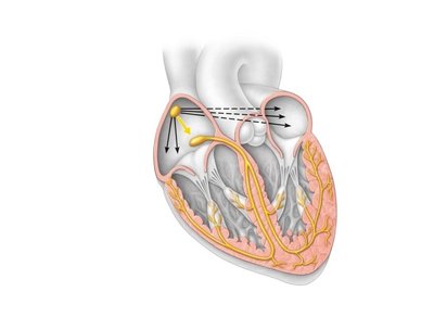
Sinoatrial (SA) Node: The pacemaker generates impulses about 75 times per minute (sinus rhythm).
Atrioventricular (AV) Node: Delays impulses by 0.1 second, allowing atrial contraction to finish before ventricular contraction begins.
AV Bundle (Bundle of His): The only electrical connection between atria and ventricles.
Right and Left Bundle Branches: Carry impulses toward the apex of the heart.
Purkinje Fibers: Complete the pathway into the apex and ventricular walls.
Extrinsic Innervation of the Heart
The autonomic nervous system (ANS) modifies the heartbeat:
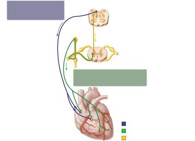
Cardioacceleratory Center: Located in the medulla oblongata, it increases heart rate and force via sympathetic neurons.
Cardioinhibitory Center: Decreases heart rate via parasympathetic fibers in the vagus nerve.
Electrocardiography (ECG/EKG)
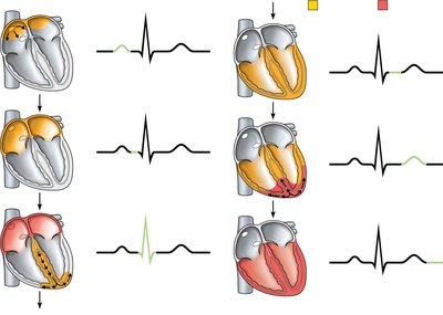
An ECG is a composite of all action potentials generated by nodal and contractile cells at a given time. It consists of three main waves:
P Wave: Depolarization of the SA node (atrial depolarization).
QRS Complex: Ventricular depolarization.
T Wave: Ventricular repolarization.
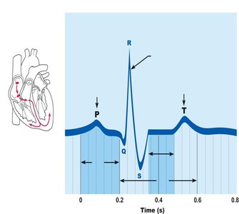
The ECG also includes intervals and segments that reflect the timing of electrical events in the heart.
Homeostatic Imbalances of Cardiac Conduction
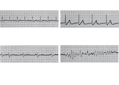
Defects in the conduction system can lead to:
Arrhythmias: Irregular heart rhythms.
Fibrillation: Rapid, irregular contractions that are ineffective for pumping blood.
Ectopic Focus: Abnormal pacemaker activity.
Heart Block: Impaired conduction between atria and ventricles.
Heart Sounds
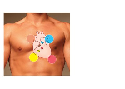
The heart produces two main sounds (lub-dup) associated with valve closure:
First Sound: AV valves close, marking the beginning of systole.
Second Sound: SL valves close, marking the beginning of diastole.
Heart Murmurs: Abnormal sounds, often indicative of valve problems.
The Cardiac Cycle
The cardiac cycle includes all events associated with blood flow during one heartbeat:
Systole: Contraction phase.
Diastole: Relaxation phase.
Phases of the Cardiac Cycle
Ventricular Filling: Occurs in late diastole; AV valves open, and most blood flows passively into ventricles. Atrial systole delivers the remaining blood.
Ventricular Systole: QRS complex signals ventricular depolarization and contraction. AV valves close (first heart sound), and SL valves open during ejection phase.
Diastole: T wave signals ventricular repolarization and relaxation. SL valves close (second heart sound), and AV valves reopen to start the next cycle.
Cardiac Output (CO)
Cardiac output is the volume of blood pumped by each ventricle per minute:
Formula:
Heart Rate (HR): Beats per minute.
Stroke Volume (SV): Volume of blood pumped per beat.
At rest:
Cardiac Reserve: Difference between resting and maximal CO.
Regulation of Stroke Volume
Stroke volume is determined by:
Formula:
Preload: Degree of stretch before contraction (Frank-Starling law).
Contractility: Strength of contraction at a given muscle length.
Afterload: Pressure that must be overcome to eject blood.
Regulation of Heart Rate
Heart rate is influenced by:
Positive Chronotropic Factors: Increase heart rate (e.g., sympathetic stimulation, hormones).
Negative Chronotropic Factors: Decrease heart rate (e.g., parasympathetic stimulation).
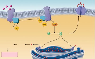
The autonomic nervous system regulates heart rate via sympathetic (norepinephrine) and parasympathetic (acetylcholine) pathways. The heart at rest exhibits vagal tone (parasympathetic dominance).
Chemical Regulation of Heart Rate
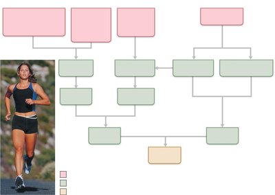
Hormones and ion concentrations affect heart rate:
Epinephrine: Enhances heart rate and contractility.
Thyroxine: Increases heart rate and potentiates catecholamine effects.
Ca2+ and K+: Must be maintained for normal function.
Other Factors Influencing Heart Rate
Age: Heart rate decreases with age.
Gender: Females typically have higher heart rates.
Exercise: Increases heart rate and cardiac output.
Body Temperature: Higher temperatures increase heart rate.
Homeostatic Imbalances
Tachycardia: Abnormally fast heart rate (>100 bpm).
Bradycardia: Abnormally slow heart rate (<60 bpm).
Congestive Heart Failure (CHF): Progressive condition where cardiac output is insufficient for tissue needs.
Developmental Aspects of the Heart
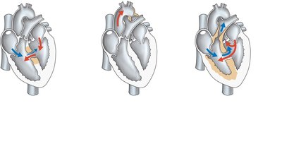
Congenital heart defects can lead to mixing of systemic and pulmonary blood or increased workload due to narrowed valves or vessels.
Age-Related Changes Affecting the Heart
Sclerosis and Thickening of Valve Flaps: Can impair valve function.
Decline in Cardiac Reserve: Reduced ability to increase cardiac output.
Fibrosis of Cardiac Muscle: Decreases contractility.
Atherosclerosis: Hardening and narrowing of arteries, increasing risk of heart disease.
