 뒤로
뒤로The Cellular Level of Organization: Structure, Function, and Genetic Control
스터디 가이드 - 스마트 노트
 자료에 맞춘 맞춤형 노트, 핵심 정의, 예시, 맥락을 확장해 제공합니다.
The Cellular Level of Organization
The Cell Theory
The cell theory is a fundamental concept in biology, establishing the cell as the basic unit of life. Developed in the 19th century, it provides the foundation for understanding the structure and function of all living organisms.
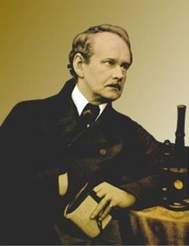
Matthias Schleiden (1838): Concluded that all plants are composed of cells.
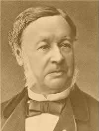
Theodor Schwann (1839): Concluded that all animals are composed of cells.
Rudolph Virchow (1855): Determined that cells come only from other cells.
Principles of Cell Theory:
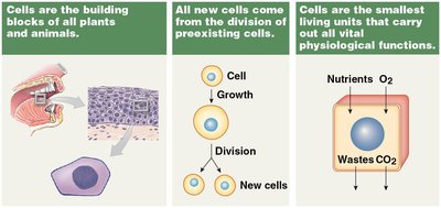
Cells are the building blocks of all plants and animals.
All new cells come from the division of preexisting cells.
Cells are the smallest living units that perform all vital physiological functions.
Cell Differentiation
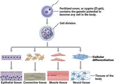
Cell differentiation is the process by which cells become specialized to perform specific functions. This process begins with a fertilized ovum and leads to the formation of various tissue types in the body.
Cellular differentiation: Gradual specialization of cells as they divide and mature.
Major tissue types: Epithelial, connective, muscle, and neural tissues.
Cell Structure and Function
Body Fluid Distribution
Cells are surrounded by extracellular fluid, while the fluid inside the cell is called intracellular fluid or cytosol. The plasma membrane separates these two environments, maintaining cellular integrity.
Extracellular fluid: Watery medium surrounding cells.
Intracellular fluid (cytosol): Fluid within the cell.
Plasma membrane: Separates cytoplasm from extracellular fluid.
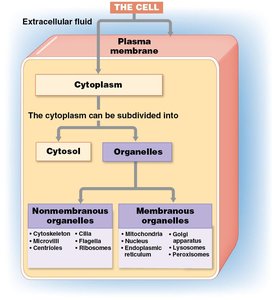
Cytoplasm: Contains cytosol and organelles.
Organelles
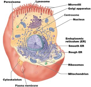
Organelles are specialized structures within cells, divided into membranous and nonmembranous types. Each organelle performs distinct functions essential for cell survival.
Nonmembranous organelles: Not enclosed by membranes; in direct contact with cytosol (e.g., cytoskeleton, ribosomes).
Membranous organelles: Enclosed in a phospholipid membrane; isolated from cytosol (e.g., mitochondria, Golgi apparatus).
Peroxisome
Peroxisomes are vesicles containing degradative enzymes. They break down organic compounds and neutralize toxic substances.
Structure: Vesicles with degradative enzymes.
Function: Breakdown of organic compounds; neutralization of toxins.
Lysosome
Lysosomes are vesicles containing digestive enzymes. They break down organic compounds, damaged organelles, and pathogens.
Structure: Vesicles with digestive enzymes.
Function: Digestion of organic compounds and cellular debris.
Mitochondrion
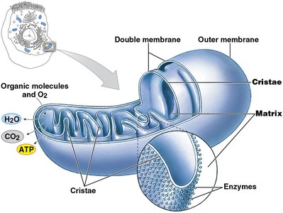
Mitochondria are double-membraned organelles responsible for producing most of the cell's ATP through aerobic metabolism.
Structure: Double membrane; inner membrane contains metabolic enzymes.
Function: Produces 95% of cellular ATP.
Cytoskeleton
The cytoskeleton provides structural support and facilitates movement of cellular structures and materials.
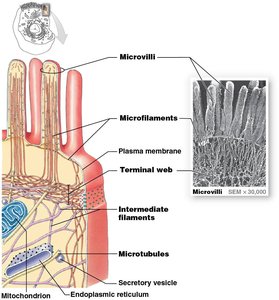
Structure: Proteins organized into filaments and tubes; includes microfilaments, intermediate filaments, and microtubules.
Function: Strengthens and supports cell; enables movement within the cell.
Plasma Membrane
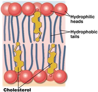
The plasma membrane is a selectively permeable barrier composed of a phospholipid bilayer, proteins, and carbohydrates.
Phospholipid bilayer: Provides structural foundation.
Proteins: Integral (embedded) and peripheral (surface-associated).
Glycocalyx: Carbohydrate-rich layer for cell recognition and binding.
Cytoskeleton Components
The cytoskeleton consists of microfilaments, intermediate filaments, and microtubules, each with distinct structural and functional roles.
Microfilaments: Actin protein, <6nm diameter.
Intermediate filaments: 7-11nm diameter.
Microtubules: Tubulin protein, ~25nm diameter.
Ribosomes
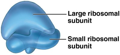
Ribosomes are responsible for protein synthesis and consist of two subunits containing proteins and ribosomal RNA (rRNA).
Free ribosomes: Located in cytoplasm; synthesize proteins for use within the cell.
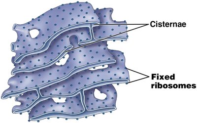
Bound ribosomes: Attached to rough ER; synthesize proteins for export.
Endoplasmic Reticulum (ER)
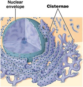
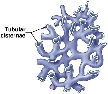
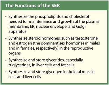
The ER is a network of membranes continuous with the nuclear envelope, involved in synthesis and storage of proteins, lipids, and carbohydrates.
Smooth ER (SER): Lacks ribosomes; synthesizes lipids and carbohydrates.
Rough ER (RER): Has attached ribosomes; modifies and exports proteins.
Golgi Apparatus
The Golgi apparatus functions as a packaging center, modifying and packaging secretions and enzymes for release or use within the cell.
Renews/modifies plasma membrane.
Packages secretions for exocytosis.
Packages enzymes for lysosomes.
Mitochondria and ATP Production
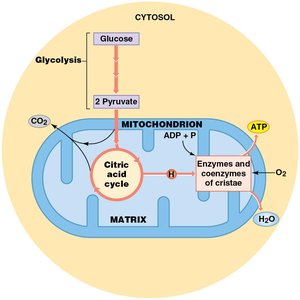
Mitochondria are the powerhouses of the cell, producing ATP through aerobic metabolism. The number of mitochondria varies by cell type and energy requirements.
Contain their own DNA and ribosomes.
ATP production steps:
Glycolysis in cytosol: 1 glucose → 2 pyruvate.
Pyruvate enters mitochondria; CO2 removed; enters citric acid cycle.
Enzymes and coenzymes catalyze ATP formation from ADP.
ATP leaves mitochondrion.
The Nucleus and Genetic Control
Nucleus
The nucleus is the control center for cellular homeostasis, directing synthesis of proteins and storing genetic information.
Nuclear envelope: Double membrane separating nucleus from cytoplasm.
Nuclear pores: Allow communication between nucleus and cytoplasm.
Nucleoplasm: Fluid contents of nucleus, containing structural filaments, ions, enzymes, nucleotides, RNA, and DNA.
DNA Organization
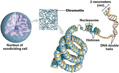
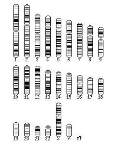
DNA in the nucleus stores instructions for protein synthesis. It is organized as chromatin in nondividing cells and chromosomes in dividing cells.
Chromatin: Loosely coiled DNA.
Chromosomes: Tightly coiled DNA during cell division.
Nucleosomes: DNA wrapped around histone proteins.
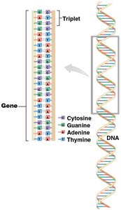
Genetic Code and Protein Synthesis
The genetic code is stored in the sequence of DNA base pairs. Protein synthesis involves transcription and translation, utilizing DNA, enzymes, and three types of RNA.
DNA bases: Adenine (A), Thymine (T), Cytosine (C), Guanine (G).
Triplet code: Sequence of three bases specifies an amino acid.
Gene: Functional unit of heredity; contains DNA to produce a specific protein.
Steps in Protein Synthesis
Protein synthesis is a multi-step process involving gene activation, transcription, and translation.
Gene activation: Histones removed, DNA uncoils.
DNA strands separate.
Enzymes assemble nucleotides into mRNA (transcription).
mRNA leaves nucleus through nuclear pores.
At ribosome, mRNA codons bind to tRNA anticodons (translation).
tRNA carries specific amino acids.
rRNA strings amino acids together to form proteins.
Plasma Membrane Permeability and Transport
Permeability
The plasma membrane is selectively permeable, allowing certain substances to pass while restricting others. Permeability depends on size, shape, lipid solubility, and electrical charge.
Freely permeable: Any substance can pass (not found in living cells).
Selectively permeable: Some substances cross.
Impermeable: No substances can pass (not found in living cells).
Types of Membrane Transport
Membrane transport is classified as passive (no ATP required) or active (requires ATP).
Passive transport: Diffusion, carrier-mediated transport.
Active transport: Vesicular transport, carrier-mediated transport.
Diffusion
Diffusion is the passive movement of substances from higher to lower concentration, driven by concentration gradients.
Factors influencing diffusion: Distance, molecule size, temperature, concentration gradient, electrical forces.
Osmosis
Osmosis is the diffusion of water across a selectively permeable membrane, moving toward higher solute concentration.
Osmotic pressure: Force of water moving into a solution.
Hydrostatic pressure: Fluid force opposing osmotic flow.
Osmolarity and Tonicity
Osmolarity is the total solute concentration in a solution, while tonicity describes the effect of osmotic solutions on cell volume.
Isotonic solution: Same osmolarity as intracellular fluid; no net water movement.
Hypertonic solution: Higher osmolarity; cell shrinks.
Hypotonic solution: Lower osmolarity; cell swells.
Carrier-Mediated and Active Transport
Carrier-mediated transport involves proteins that move substances across membranes. Active transport requires ATP and can move substances against concentration gradients.
Ion pumps: Move ions like Na+, K+, Ca2+, Mg2+.
Sodium-potassium ATPase: Exchanges 3 Na+ for 2 K+.
Vesicular Transport
Vesicular transport moves materials in membranous sacs (vesicles) and requires ATP. Includes endocytosis (import) and exocytosis (export).
Endocytosis: Importing substances into vesicles.
Exocytosis: Exporting substances from vesicles to outside the cell.
Cell Life Cycle and Division
Cell Division
Cell division is essential for growth, development, and maintenance. Most cells spend their life in interphase, with division occurring through mitosis or meiosis.
Mitosis: Produces two daughter cells with 46 chromosomes each.
Meiosis: Produces sex cells with 23 chromosomes each.
DNA replication: Helicases unwind DNA; DNA polymerase assembles new strands; ligases join sections.
Tumors and Cancer
Cancer and Tumor Formation
Cancer is characterized by abnormal cell growth and division, often due to permanent DNA mutations. Tumors may be benign or malignant, with malignant tumors capable of invading tissues and metastasizing.
Benign tumor: Cells remain within original tissue; seldom a threat.
Malignant tumor: Rapid division, angiogenesis, invasion, and metastasis.
Cancer cells: Disrupt normal function, compete for resources, and may produce abnormal substances.
Type | Characteristics |
|---|---|
Benign | Localized, non-invasive, removable |
Malignant | Invasive, rapid growth, metastasis |
Additional info: Cancer is most common in tissues with actively dividing cells, such as skin and intestinal lining.
