 뒤로
Ch.12 Part 2
스터디 가이드 - 스마트 노트
 자료에 맞춘 맞춤형 노트, 핵심 정의, 예시, 맥락을 확장해 제공합니다.
The Diencephalon
Overview of the Diencephalon
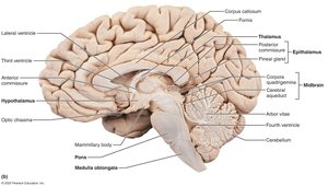
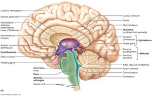
The diencephalon is a central region of the brain composed of three paired gray-matter structures: the thalamus, hypothalamus, and epithalamus. These structures enclose the third ventricle and play critical roles in sensory processing, homeostasis, and endocrine regulation.
Thalamus: Acts as a relay station for sensory and motor signals to the cerebral cortex.
Hypothalamus: Main visceral control center, regulating homeostasis and endocrine functions.
Epithalamus: Contains the pineal gland, which regulates sleep-wake cycles.
Thalamus
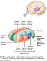
The thalamus consists of bilateral egg-shaped nuclei forming the superolateral walls of the third ventricle. It makes up 80% of the diencephalon and is connected by the interthalamic adhesion. The thalamus contains several nuclei, each named for its location, which project and receive fibers from the cerebral cortex.
Main function: Relay station for information entering the cortex.
Processes: Sorts, edits, and relays ascending input, including impulses for emotion, visceral function, motor control, memory, and sensory integration.
Role: Mediates sensation, motor activities, cortical arousal, learning, and memory.
Hypothalamus
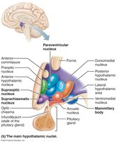
The hypothalamus is located below the thalamus, forming the cap over the brainstem and the inferolateral walls of the third ventricle. It contains important nuclei such as the mammillary bodies (olfactory relay stations) and the infundibulum (stalk connecting to the pituitary gland).
Main visceral control center: Vital to homeostasis.
Controls autonomic nervous system: Regulates blood pressure, heart rate, digestive tract motility, and pupil size.
Initiates physical responses to emotions: Perceives pleasure, fear, rage, biological rhythms, and drives.
Regulates: Body temperature, hunger, satiety, water balance, thirst, sleep-wake cycles, and sets the biological clock.
Endocrine functions: Controls secretions of the anterior pituitary gland and produces posterior pituitary hormones.
Epithalamus
The epithalamus is the most dorsal portion of the diencephalon, forming the roof of the third ventricle. It contains the pineal gland, which secretes melatonin to regulate the sleep-wake cycle.
The Brain Stem
Overview of the Brain Stem
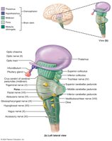
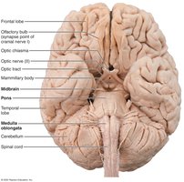
The brain stem consists of three regions: the midbrain, pons, and medulla oblongata. It is structurally similar to the spinal cord but contains nuclei embedded in white matter. The brain stem controls automatic behaviors necessary for survival and connects higher and lower neural centers. Nuclei in the brain stem are associated with 10 of the 12 pairs of cranial nerves.
Midbrain
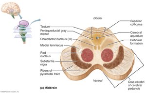
The midbrain is located between the diencephalon and pons. It contains the cerebral peduncles (ventral bulges with pyramidal motor tracts) and the cerebral aqueduct (channel connecting the third and fourth ventricles).
Corpora quadrigemina: Paired dorsal protrusions, including superior colliculi (visual reflex centers) and inferior colliculi (auditory relay centers).
Substantia nigra: Functionally linked to basal nuclei; degeneration leads to Parkinson’s disease.
Red nucleus: Relay nuclei for descending limb flexion motor pathways; part of the reticular formation.
Pons
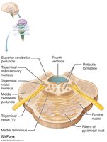
The pons is located between the midbrain and medulla oblongata, separated from the cerebellum by the fourth ventricle. It is composed of conduction tracts, including longitudinal fibers (connecting higher brain centers and spinal cord) and transversal/dorsal fibers (relaying impulses between motor cortex and cerebellum).
Origin of cranial nerves: Some nuclei play roles in the reticular formation and help maintain normal breathing rhythm.
Medulla Oblongata
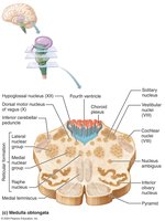
The medulla oblongata blends into the spinal cord at the foramen magnum and contains the fourth ventricle. It is a continuation of the central canal of the spinal cord and forms the ventral wall with the pons. The medulla contains the choroid plexus, which forms cerebrospinal fluid.
Pyramids: Ventral ridges formed by pyramidal tracts from the motor cortex.
Decussation of the pyramids: Point where tracts cross to the opposite side of the body.
Olives: Swellings caused by inferior olivary nuclei, relaying stretch information to the cerebellum.
Cranial nerves: Vestibular and cochlear nuclei (equilibrium), nucleus cuneatus and gracilis (sensory relay).
Autonomic reflex center: Cardiovascular center (heart rate and blood pressure), respiratory centers (breathing rhythm), and centers for vomiting, hiccupping, swallowing, coughing, and sneezing.
Spinal Cord: Gross Anatomy and Protection
Overview and Functions
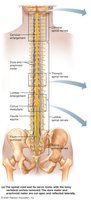
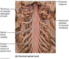
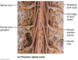
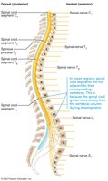
The spinal cord is enclosed in the vertebral column, beginning at the foramen magnum and ending at the L1 or L2 vertebra. It provides two-way communication between the brain and body and serves as a major reflex center.
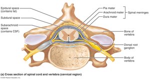
Protected by: Bone, meninges, and cerebrospinal fluid (CSF).
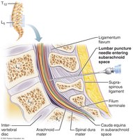
Spinal dura mater: One layer thick, not attached to vertebrae.
Epidural space: Cushion of fat and veins between vertebrae and dura mater.
CSF: Fills subarachnoid space between arachnoid and pia maters.
Dural and arachnoid membranes: Extend to sacrum, site of lumbar puncture.
Spinal Cord Structure
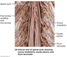
Conus medullaris: Cone-shaped terminal end of the spinal cord.
Filum terminale: Fibrous extension anchoring the cord to the coccyx.
Denticulate ligaments: Extensions of pia mater securing the cord to dura mater.
Cervical and lumbar enlargements: Areas where nerves serving limbs arise.
Spinal nerves: 31 pairs attach to the cord by paired roots.
Cauda equina: Collection of nerve roots at the inferior end of the vertebral canal.
Spinal Cord Cross-Sectional Anatomy
Gray and White Matter
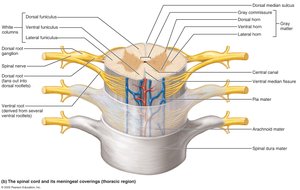
The spinal cord is divided into right and left halves by the ventral (anterior) median fissure and dorsal (posterior) median sulcus. Gray matter is located in the core, surrounded by white matter. The central canal runs the length of the cord and is filled with CSF.
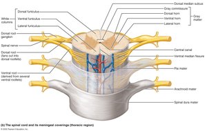
Gray matter: Resembles a butterfly or letter "H" in cross section. Contains dorsal horns (interneurons for sensory input), ventral horns (motor neurons), and lateral horns (sympathetic neurons in thoracic/lumbar regions).
Gray commissure: Bridge connecting gray matter masses, enclosing the central canal.
Ventral roots: Motor neuron axons exiting the cord.
Dorsal roots: Sensory input to the cord; dorsal root ganglia contain sensory neuron cell bodies.
Spinal nerves: Formed by fusion of dorsal and ventral roots.
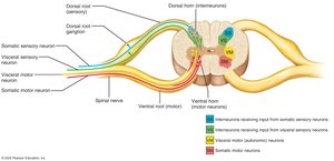
Gray matter groups: Somatic sensory (SS), visceral sensory (VS), visceral motor (VM), and somatic motor (SM).
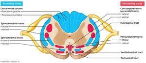
White matter: Myelinated and nonmyelinated fibers allow communication between spinal cord parts and between the cord and brain. Fibers run ascending (sensory), descending (motor), and transverse (commissural).
White columns (funiculi): Dorsal, lateral, and ventral columns on each side.
Spinal tracts: Composed of axons with similar destinations and functions.
Spinal Cord Trauma and Disorders
Types of Trauma
Paresthesias: Sensory function loss due to dorsal root or sensory tract damage.
Paralysis: Motor function loss due to ventral root or ventral horn cell damage.
Flaccid paralysis: Severe damage to ventral root/horn; no voluntary/involuntary control, muscle atrophy.
Spastic paralysis: Damage to upper motor neurons; reflex activity remains, no voluntary control, muscles may shorten permanently.
Transection: Cross-sectioning of the cord results in total motor and sensory loss below the cut. Paraplegia (T1–L1), quadriplegia (cervical region), spinal shock (transient loss caudal to lesion).
Protection of the Brain: Meninges
Meninges Structure and Function
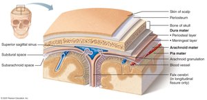
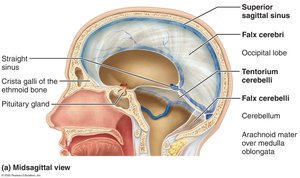
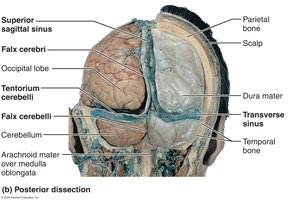
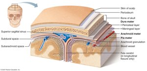
The meninges are three connective tissue membranes that cover and protect the CNS, protect blood vessels, contain CSF, and form partitions in the skull. The three layers are dura mater, arachnoid mater, and pia mater.
Dura mater: Strongest meninx, two layers in the brain (periosteal and meningeal), forms dural venous sinuses and dural septa (falx cerebri, falx cerebelli, tentorium cerebelli).
Arachnoid mater: Middle layer with spiderweb-like extensions, separated from dura by subdural space, subarachnoid space contains CSF and blood vessels, arachnoid granulations reabsorb CSF.
Pia mater: Delicate layer clinging tightly to the brain, contains many blood vessels.
Clinical Note: Meningitis
Meningitis is the inflammation of the meninges, which may spread to the CNS and cause encephalitis (inflammation of the brain). Diagnosis is typically made by observing microbes in a CSF sample obtained via lumbar puncture.
----------------------------------------
