 뒤로
The Central Nervous System: Structure and Function
스터디 가이드 - 스마트 노트
 자료에 맞춘 맞춤형 노트, 핵심 정의, 예시, 맥락을 확장해 제공합니다.
The Central Nervous System (CNS)
Introduction to the CNS
The central nervous system (CNS) is composed of the brain and spinal cord, serving as the main integration and control center of the body. It receives sensory information from the peripheral nervous system, processes and interprets this information, and coordinates motor responses that travel back through descending pathways to the periphery.
Sensory (afferent) pathways: Carry information from the body to the CNS.
Motor (efferent) pathways: Transmit commands from the CNS to muscles and glands.
Major Divisions of the Brain
Four Main Areas
The brain is divided into four major regions, each with distinct functions:
Cerebrum: Responsible for conscious thought, reasoning, memory, sensory interpretation, and voluntary movement. It is the largest region.
Diencephalon: Contains the thalamus, hypothalamus, and epithalamus; involved in sensory relay and regulation of homeostasis.
Brainstem: Connects the brain to the spinal cord and controls essential automatic functions. Includes the midbrain, pons, and medulla oblongata.
Cerebellum: Coordinates movement, posture, and balance.
The Cerebrum
Structure and Function
The cerebrum is the largest region of the brain, divided into right and left hemispheres. It is responsible for conscious awareness, reasoning, memory, voluntary movement, and sensory interpretation. The outer layer, the cerebral cortex, is composed of gray matter (neuron cell bodies) and is involved in processing and integration. The deeper region contains white matter (myelinated axons) for transmitting information between brain areas.
Surface Anatomy of the Cerebrum
The surface of the cerebrum features:
Gyri: Raised folds or ridges.
Sulci: Shallow grooves separating gyri.
Fissures: Deep grooves separating major regions.
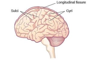
Importance of Gyri and Sulci
The folds created by gyri and sulci increase the surface area of the cerebral cortex, allowing more neurons to fit within the skull and enhancing the brain's processing capacity.
Gyri: Ridges that increase surface area.
Sulci: Grooves that separate gyri.
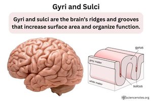
Major Fissures of the Cerebrum
Key fissures include:
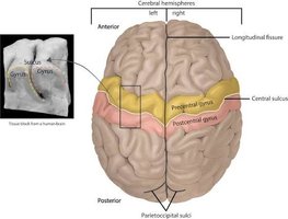
Longitudinal fissure: Separates the right and left cerebral hemispheres.
Transverse cerebral fissure: Separates the cerebrum from the cerebellum.
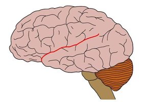
Sylvain fissure (lateral sulcus): Separates the temporal lobe from the frontal and parietal lobes.
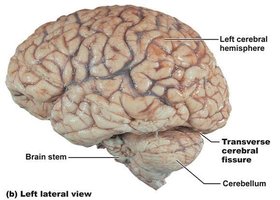
The Central Sulcus
The central sulcus is a prominent groove separating the frontal lobe from the parietal lobe. The precentral gyrus (frontal side) is involved in motor control, while the postcentral gyrus (parietal side) processes sensory information.
The Four Major Lobes of the Cerebrum
Lobe Functions
Each cerebral hemisphere contains four major lobes:
Frontal Lobe: Voluntary motor control, planning, judgment, aspects of speech production.
Parietal Lobe: Processes touch, pain, temperature, and body position.
Temporal Lobe: Hearing, memory, aspects of language comprehension, olfaction.
Occipital Lobe: Processes visual information.
Functional Areas of the Cerebral Cortex
Motor, Sensory, and Association Areas
The cerebral cortex contains three major functional areas:
Motor Area: Controls voluntary skeletal muscle movement, primarily in the frontal lobe. Includes Broca’s area for motor speech.
Sensory Area: Receives and interprets sensory information. Touch is mainly in the parietal lobe; vision in the occipital lobe; hearing and olfaction in the temporal lobe.
Association Areas: Integrate information from different regions, involved in reasoning, memory, language, and decision making. Includes Wernicke’s area for language comprehension.
Premotor Cortex, Primary Motor Cortex, and Somatosensory Cortex
These important functional areas lie on opposite sides of the central sulcus:
Premotor Cortex: Plans and prepares voluntary movements; located anterior to the primary motor cortex.
Primary Motor Cortex: Initiates voluntary skeletal muscle movement; located in the precentral gyrus.
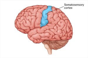
Primary Somatosensory Cortex: Receives and processes information about touch, pressure, pain, temperature, and body position; located in the postcentral gyrus.
Supplemental Motor Cortex: Organizes and coordinates movements involving both sides of the body.
Simple Summary
Premotor Cortex: Plans and prepares voluntary movements.
Supplementary Motor Cortex: Organizes and coordinates sequences of movement.
Primary Motor Cortex: Sends motor commands to execute voluntary movements.
Primary Somatosensory Cortex: Receives and processes sensory information from the body.
The Corpus Callosum
Structure and Function
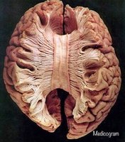
The corpus callosum is a large bundle of white matter fibers connecting the right and left cerebral hemispheres, allowing communication between them. It helps coordinate sensory, motor, and cognitive abilities.
White matter: Composed of myelinated fibers.
Example: Coordinated use of both hands requires communication between hemispheres.
The Diencephalon
Structure and Function
The diencephalon is located between the cerebral hemispheres, superior to the midbrain. It serves as a major sensory relay and regulatory center, directing sensory information to the cerebral cortex and helping maintain homeostasis.
Thalamus: Major sensory relay station, directing most sensory information (except smell) to the appropriate areas of the cerebral cortex.
Hypothalamus: Maintains homeostasis by regulating body temperature, hunger, thirst, autonomic nervous system activity, and hormone secretion.
Epithalamus: Contains the pineal gland, which produces melatonin and helps regulate the sleep-wake cycle.
Key Point: The thalamus relays sensory information and the hypothalamus regulates the body’s internal environment.
