 뒤로
뒤로The Digestive System: Structure, Function, and Clinical Relevance
스터디 가이드 - 스마트 노트
 자료에 맞춘 맞춤형 노트, 핵심 정의, 예시, 맥락을 확장해 제공합니다.
The Digestive System
Overview and Functions
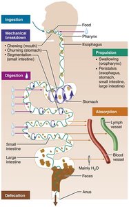
The digestive system is essential for life, responsible for converting food into absorbable nutrients and eliminating indigestible residues. It consists of a series of organs forming the alimentary canal and accessory organs that aid in digestion.
Ingestion: Intake of food into the mouth.
Propulsion: Movement of food through the digestive tract, primarily via peristalsis (alternating waves of contraction and relaxation).
Mechanical Breakdown: Physical breakdown of food (chewing, churning, segmentation).
Digestion: Chemical breakdown of food by enzymes into smaller molecules.
Absorption: Transport of digested nutrients into blood or lymph.
Defecation: Elimination of indigestible substances as feces.
General Anatomy of the Digestive System
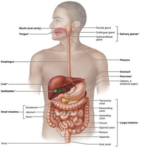
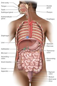
The digestive system is divided into two main groups: the alimentary canal and accessory digestive organs.
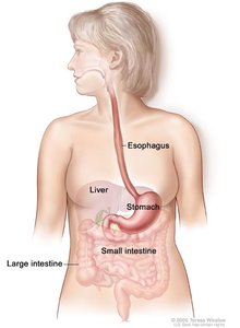
Alimentary Canal: A continuous muscular tube (~30 ft in cadaver) from mouth to anus. Includes mouth, pharynx, esophagus, stomach, small intestine, large intestine, and anus.
Accessory Organs: Teeth, tongue, gallbladder, salivary glands, liver, and pancreas. These organs secrete digestive fluids into the GI tract via ducts.
Histological Organization of the Digestive Tract
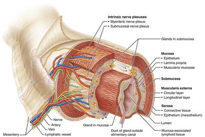
The wall of the alimentary canal consists of four main layers (tunics), each with specialized functions:
Mucosa: Innermost layer; secretes mucus, digestive enzymes, and hormones; absorbs end products; protects against infection. Composed of epithelium, lamina propria, and muscularis mucosae. Contains mucosa-associated lymphatic tissue (MALT).
Submucosa: Areolar connective tissue with blood and lymphatic vessels, lymphoid follicles, and nerve fibers. Contains abundant elastic fibers.
Muscularis Externa: Responsible for segmentation and peristalsis; consists of inner circular and outer longitudinal layers of smooth muscle. Circular layer forms sphincters.
Serosa: Outermost layer; areolar connective tissue covered by mesothelium. In organs outside the abdominopelvic cavity, serosa is replaced by adventitia.
Enteric Nervous System (ENS)
Structure and Function
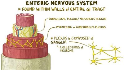
The enteric nervous system (ENS) is a complex network of neurons embedded in the walls of the GI tract, primarily within the submucosa and muscularis externa. It coordinates muscle contractions and regulates secretions to maintain optimal pH and biochemical conditions for enzyme function.
ENS can function independently of the central nervous system (CNS) but also communicates with it.
It links emotional and cognitive brain centers with peripheral intestinal functions, influenced by the gut microbiome.
Mesenteries and Omentum
Support and Protection
Mesenteries are connective tissue sheets that loosely suspend the intestines from the abdominal wall, preventing twisting and providing a passage for blood vessels, nerves, and lymphatics. The omentum is a fatty blanket (greater and lesser omentum) that covers the intestines and stores adipose tissue.
The Mouth and Associated Structures
Anatomy of the Mouth
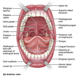
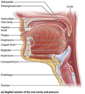
The mouth is the entry point for food and is associated with the tongue, teeth, and salivary glands. The palate forms the roof of the mouth and consists of the hard palate (bony) and soft palate (muscular). The uvula and palatine tonsils are also present.
Tongue and Tonsils
The tongue manipulates food, aids in speech, and is covered with nonkeratinized stratified squamous epithelium. It contains various papillae (filiform, fungiform) and is associated with the lingual and palatine tonsils.
Teeth (Dentition)
Teeth (dentition) are responsible for mastication (chewing). Adults typically have 32 teeth: incisors, canines, premolars, and molars. Each tooth consists of enamel (outer), dentin (middle), and pulp (center).
Salivary Glands
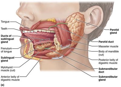
Salivary glands secrete saliva, which cleanses the mouth, dissolves food chemicals, moistens food, and begins starch digestion via amylase. Major glands include the parotid, submandibular, and sublingual glands.
Pharynx and Esophagus
Pharynx
The pharynx is a muscular funnel where the respiratory and digestive tracts intersect. It contains pharyngeal constrictors that force food downward during swallowing.
Esophagus
The esophagus is a muscular tube that connects the pharynx to the stomach. It passes through the diaphragm at the esophageal hiatus and is closed by the lower esophageal sphincter when not swallowing. The esophagus has all four GI tract tunics, with mucosa transitioning from nonkeratinized stratified squamous epithelium to simple columnar at the stomach. The submucosa contains mucus-secreting glands, and the muscularis externa transitions from skeletal to smooth muscle.
Clinical Homeostatic Imbalances
Ankyloglossia ("Fused Tongue")
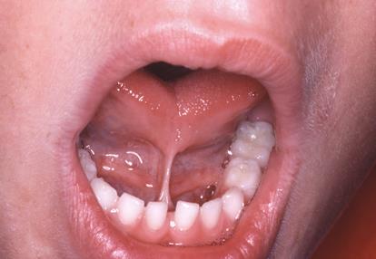
Ankyloglossia is a congenital condition characterized by a short lingual frenulum, restricting tongue movement. It may interfere with speech and breastfeeding and is treated surgically.
Heartburn and GERD
Heartburn is a burning pain caused by stomach acid entering the esophagus, often the first symptom of gastroesophageal reflux disease (GERD). GERD can result from excess food, obesity, pregnancy, running, or hiatal hernia. Treatment includes lifestyle changes, dietary modifications, and medications.
Summary Table: Major Digestive Enzymes
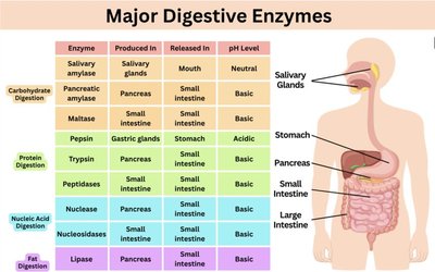
The following table summarizes the major digestive enzymes, their sources, release sites, and optimal pH levels:
Enzyme | Produced In | Released In | pH Level |
|---|---|---|---|
Salivary amylase | Salivary glands | Mouth | Neutral |
Pancreatic amylase | Pancreas | Small intestine | Basic |
Maltase | Small intestine | Small intestine | Basic |
Pepsin | Gastric glands | Stomach | Acidic |
Trypsin | Pancreas | Small intestine | Basic |
Peptidases | Small intestine | Small intestine | Basic |
Nuclease | Pancreas | Small intestine | Basic |
Nucleosidases | Small intestine | Small intestine | Basic |
Lipase | Pancreas | Small intestine | Basic |
Additional info: The digestive system also includes the liver, gallbladder, and pancreas, which will be covered in detail in subsequent sections. The gut microbiome is increasingly recognized for its role in regulating digestive and immune functions.
