 뒤로
The Endocrine System: Structure, Function, and Clinical Relevance
스터디 가이드 - 스마트 노트
 자료에 맞춘 맞춤형 노트, 핵심 정의, 예시, 맥락을 확장해 제공합니다.
The Endocrine System
Overview of the Endocrine System
The endocrine system is a network of glands that secrete chemical signals called hormones into the bloodstream. These hormones bind to specific receptors on target cells, stimulating them to respond in particular ways. The endocrine system regulates growth, metabolism, reproduction, and homeostasis.
Hormones: Chemical messengers released by glands.
Target cells: Cells with receptors for specific hormones.
Regulation: Hormone secretion is controlled by feedback mechanisms involving the hypothalamus and pituitary gland.
Pituitary Gland
Anterior Pituitary Gland
The anterior pituitary gland produces several key hormones that regulate growth, metabolism, and reproduction.
Growth Hormone (GH, Somatotropin): Promotes growth by stimulating cells to produce insulin-like growth factors (IGFs), increases amino acid transport, and stimulates cell division. Regulated by GHRH (stimulates) and GHIH/somatostatin (inhibits) from the hypothalamus.
Thyroid Stimulating Hormone (TSH, Thyrotropin): Stimulates the thyroid gland to produce thyroxine (T4) and triiodothyronine (T3).
Adrenocorticotropic Hormone (ACTH): Stimulates the adrenal gland to secrete cortisol, which increases the use of fat and amino acids for energy.
Prolactin: Induces lactation in mammary glands; regulated by PRH from the hypothalamus and influenced by estrogen and progesterone.
Follicle Stimulating Hormone (FSH) & Luteinizing Hormone (LH): Stimulate gonads to produce testosterone (males) and estrogen (females); regulated by GnRH from the hypothalamus.
Posterior Pituitary Gland
The posterior pituitary gland stores and releases hormones synthesized in the hypothalamus.
Antidiuretic Hormone (ADH, Vasopressin): Acts on kidney tubules to retain water.
Oxytocin: Initiates uterine muscle contraction and is involved in lactation.
Note: The posterior pituitary does not synthesize hormones; it stores them for release.
Thyroid Gland
Structure and Function
The thyroid gland is located on the front and sides of the trachea below the larynx. It consists of two lobes connected by the isthmus. The structural unit is the thyroid follicle, which produces T3 and T4 hormones. Iodine is essential for their synthesis.
T3 (Triiodothyronine): Contains three iodine atoms.
T4 (Thyroxine): Contains four iodine atoms.
Calcitonin: Produced by parafollicular cells; decreases reabsorption of calcium and phosphate from bone to blood.
T3 and T4 regulate energy production, protein synthesis, and metabolic rate, affecting growth, mental development, and reproductive system maturation.
Regulation of Thyroid Hormones
Stimulated by TSH from the anterior pituitary gland.
Decreased metabolic rate triggers increased thyroid hormone secretion.
Calcitonin
Decreases blood calcium and phosphate levels by inhibiting their release from bones.
Important for bone growth during childhood.
Stimulated by hypercalcemia (high blood calcium).
Parathyroid Gland
Structure and Function
The parathyroid glands are four small glands located on the posterior surface of the thyroid gland. They produce parathyroid hormone (PTH).
PTH: Antagonist to calcitonin; increases blood calcium by stimulating reabsorption from bones, kidneys, and intestines.
Stimulates activation of vitamin D, increasing calcium absorption in the intestines.
Stimulated by hypocalcemia (low blood calcium); inhibited by hypercalcemia.
Calcium is essential for blood clotting, bone formation, neuron and muscle contraction, and electrical conductivity.
Pancreas
Structure and Function
The pancreas is both an exocrine and endocrine gland. The endocrine portion consists of the Islets of Langerhans, which contain:
Alpha cells: Produce glucagon, stimulating glycogen breakdown in the liver.
Beta cells: Produce insulin, increasing glucose uptake by cells.
Delta cells: Produce somatostatin, inhibiting hormone secretion and promoting glycogen synthesis.
Insulin enables cells to use fatty acids and amino acids for synthesis of lipids and proteins. Secretion is stimulated by hyperglycemia (high blood glucose).
Adrenal Gland
Structure and Function
The adrenal glands are located on top of each kidney and consist of two parts:
Adrenal medulla: Secretes epinephrine and norepinephrine (catecholamines), which mimic sympathetic nervous system responses (fight or flight).
Adrenal cortex: Secretes mineralocorticoids (aldosterone), glucocorticoids (cortisol), and sex hormones.
Adrenal Medulla
Epinephrine: Increases heart rate, dilates bronchioles, stimulates glycogen breakdown, and increases fat use for energy.
Norepinephrine: Causes vasoconstriction and raises blood pressure.
Adrenal Cortex
Aldosterone: Increases sodium reabsorption and potassium excretion in kidneys, maintaining blood volume, pressure, and pH.
Cortisol: Increases fat and amino acid use for energy, conserves glucose for the brain, has anti-inflammatory effects, and is stimulated by ACTH.
Sex hormones: Estrogen and progesterone (ovaries), testosterone (testes).
Pineal Gland
Function
The pineal gland secretes melatonin, which regulates sleep cycles.
Hormones Produced by Tissues (Not Glands)
Erythropoietin: Stimulates red blood cell production in bone marrow (kidney tissue).
Atrial Natriuretic Peptide (ANP): Antagonist to aldosterone; induces sodium excretion to lower blood pressure.
Gastrin: Increases hydrochloric acid production in the stomach.
Secretin, Cholecystokinin (CCK), Motilin, GIP: Regulate digestive processes in the small intestine.
Prostaglandins (PG): Local hormones with roles in inflammation, pain, vasoconstriction, and metabolism.
Osteocalcin: Regulates glucose metabolism, muscle strength, and stress response (skeletal system).
Stress Response
Physiological Response to Stress
The body responds to stress through coordinated actions of the nervous and endocrine systems. The hypothalamus activates the sympathetic nervous system and adrenal glands, leading to the release of epinephrine, norepinephrine, and cortisol.
Increased heart rate, ventilation, blood pressure, and blood flow to muscles.
Mobilization of energy stores (glycogen, fat).
Suppression of insulin secretion and increased glucagon secretion.
Cortisol provides prolonged response, mobilizes amino acids, inhibits inflammation, and maintains blood pressure.
Aldosterone and ADH promote water and salt retention to maintain blood volume and pressure.
Thyroid Disorders
Hyposecretion and Hypersecretion
Hyposecretion: Lethargy, feeling cold, mental sluggishness, myxedema, impaired memory.
Hypersecretion: Restlessness, sweating, weight loss, irritability, increased heart rate, Grave's disease, exophthalmus.
Primary and Secondary Disorders
Primary hypothyroidism: Failure of thyroid gland or iodine deficiency.
Secondary hypothyroidism: Failure of pituitary gland or hypothalamus.
Primary hyperthyroidism: Thyroid tumor or Grave's disease.
Secondary hyperthyroidism: Increased secretion by pituitary or hypothalamus.
Goiter (enlarged thyroid) may occur in some conditions.
Hormones & Biochemistry
Classes of Hormones
Peptides: Chains of amino acids; water-soluble (e.g., insulin).
Amines: Derived from tyrosine; water-soluble (e.g., dopamine, norepinephrine, epinephrine). T3 and T4 are lipid-soluble.
Steroids: Derived from cholesterol; lipid-soluble (e.g., cortisol, aldosterone).
Synthesis and Release
Peptide hormones: Synthesized in the Golgi apparatus, stored in vesicles, released by exocytosis.
Amines: Synthesized from tyrosine, stored in chromaffin granules, released rapidly.
Steroids: Synthesized from cholesterol in the adrenal cortex, not stored, released by diffusion.
Stimuli Controlling Hormone Secretion
Neural stimuli: Nerve impulses trigger hormone release (e.g., acetylcholine stimulates adrenal medulla).
Hormonal stimuli: Hormones stimulate other glands (e.g., TSH stimulates thyroid).
Humoral stimuli: Changes in blood ion/nutrient levels (e.g., insulin, glucagon, aldosterone, calcitonin, PTH).
Action of Hormones
Mechanism of Action
Hormones bind to specific receptors on target cells.
Receptor activation initiates signal transduction via secondary messengers or gene expression.
Cellular responses include contraction, secretion, transport, protein synthesis, and metabolism.
Water-soluble hormones bind to plasma membrane receptors (G proteins, protein kinases).
Lipid-soluble hormones bind to intracellular receptors, acting as transcription factors to regulate gene expression.
Diabetes Mellitus
Types and Pathophysiology
Type I Diabetes: Absence of insulin production (juvenile onset).
Type II Diabetes: Insulin resistance; normal or elevated insulin levels (adult onset).
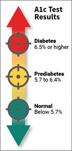
Symptoms include hyperglycemia, glycosuria, polyuria, polydipsia, polyphagia, weight loss, ketosis, and dehydration. Glycated hemoglobin (HbA1C) is used to monitor long-term glucose control.
Diabetes Mellitus vs. Diabetes Insipidus
Diabetes Mellitus: Involves increased blood glucose levels.
Diabetes Insipidus: Involves extreme water loss and electrolyte imbalance; rare.
Diabetic socks and compression socks are used to improve blood flow and prevent complications.
Lipid Soluble Hormones
Lipid-soluble hormones bind to intracellular receptors in the nucleus, acting as transcription factors to regulate gene expression and protein synthesis.
Hormone-receptor complex binds to DNA, activating or inhibiting genes.
Transcription and translation produce proteins that catalyze cellular responses.
Key Equations
HbA1C Diagnostic Criteria:
Normal: Below 5.7%
Prediabetes: 5.7% to 6.4%
Diabetes: 6.5% or higher
Additional info: Expanded explanations and clinical context were added for clarity and completeness.
