 뒤로
뒤로The Integumentary System: Structure, Function, and Clinical Aspects
스터디 가이드 - 스마트 노트
 자료에 맞춘 맞춤형 노트, 핵심 정의, 예시, 맥락을 확장해 제공합니다.
The Integumentary System
Overview
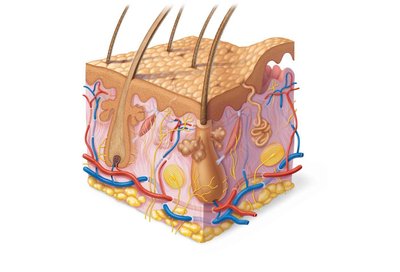
The integumentary system is the body's largest organ system, providing essential protection and sensory functions. It consists of the skin, sweat (sudoriferous) glands, oil (sebaceous) glands, hair, nails, and subcutaneous tissue. Together, these components maintain homeostasis and defend against environmental threats.
Skin: Primary barrier and sensory organ
Sweat glands: Regulate temperature and excrete wastes
Oil glands: Lubricate and protect skin and hair
Hair: Provides protection and sensory input
Nails: Protect fingertips and enhance sensation
Subcutaneous tissue: Anchors skin, absorbs shock, insulates
Skin Structure
Layers of the Skin
The skin is composed of two main layers: the epidermis and dermis. Beneath these lies the subcutaneous tissue (hypodermis), which, while not technically part of the skin, is functionally important.
Epidermis: Outermost layer, made of epithelial tissue, avascular
Dermis: Underlies epidermis, dense connective tissue, vascular
Subcutaneous tissue (Hypodermis): Mostly adipose tissue, anchors skin, provides insulation and shock absorption
The Epidermis
Cell Types
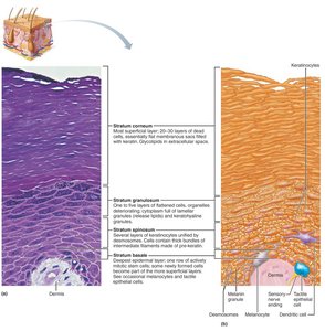
The epidermis is a keratinized stratified squamous epithelium, containing four main cell types:
Keratinocytes: Produce keratin, the protein that gives skin its strength and waterproofing
Melanocytes: Produce melanin pigment, protecting against UV radiation
Dendritic (Langerhans) cells: Immune cells that patrol for pathogens
Tactile epithelial (Merkel) cells: Sensory receptors for touch
Layers of the Epidermis
The epidermis is organized into distinct strata, each with specialized functions:
Stratum basale: Deepest layer, mitotically active stem cells, contains melanocytes
Stratum spinosum: Several layers thick, contains pre-keratin filaments, dendritic cells
Stratum granulosum: Cells flatten, keratinization begins, glycolipids for water resistance
Stratum lucidum: Only in thick skin (palms, soles), clear layer of dead keratinocytes
Stratum corneum: Outermost, 20–30 layers of dead, keratinized cells, barrier function
The Dermis
Structure and Function
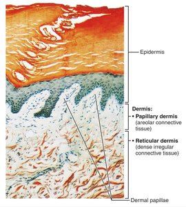
The dermis is a strong, flexible connective tissue layer containing fibroblasts, macrophages, mast cells, and white blood cells. It houses nerves, blood vessels, lymphatics, hair follicles, oil, and sweat glands.
Papillary dermis: Superficial, areolar connective tissue, contains dermal papillae with capillaries and sensory receptors
Reticular dermis: Deep, dense irregular connective tissue, provides strength, elasticity, and hydration
Dermal Modifications
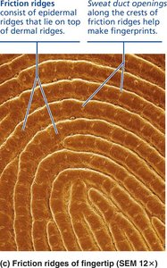
Friction ridges: Enhance grip and touch, form fingerprints
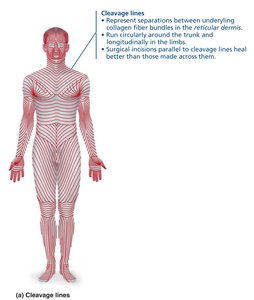
Cleavage (tension) lines: Parallel collagen fibers, important for surgical incisions
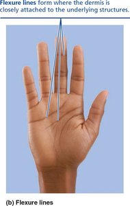
Flexure lines: Dermal folds at joints, visible on hands and feet
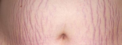
Striae (stretch marks): Dermal tears from extreme stretching
Skin Color
Pigments
Three pigments contribute to skin color:
Melanin: Produced by melanocytes, shields DNA from UV, varies in amount and form
Carotene: Yellow-orange pigment, accumulates in stratum corneum and hypodermis, can be converted to vitamin A
Hemoglobin: Red pigment in blood, gives pinkish hue to fair skin
Alterations in skin color can indicate disease (cyanosis, pallor, erythema, jaundice, bruises).
Hair
Structure and Function
Hair is composed of dead, keratinized cells and serves protective and sensory functions. It is absent from palms, soles, lips, nipples, and parts of external genitalia.
Root: Within scalp, keratinization ongoing
Shaft: Above scalp, keratinization complete
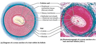
Medulla: Central core
Cortex: Surrounds medulla
Cuticle: Outermost layer
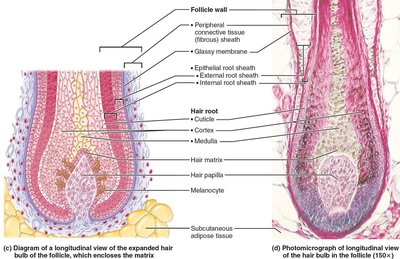
Hair Follicle
Follicle wall: Peripheral connective tissue sheath, glassy membrane, epithelial root sheath
Hair bulb: Expanded deep end, contains hair matrix (actively dividing cells)
Arrector pili: Smooth muscle causing "goose bumps"
Types and Growth
Vellus hair: Fine, pale body hair
Terminal hair: Coarse, long hair (scalp, eyebrows, axillary, pubic regions)
Growth cycles: Active and resting phases, affected by nutrition and hormones
Nails
Structure and Function
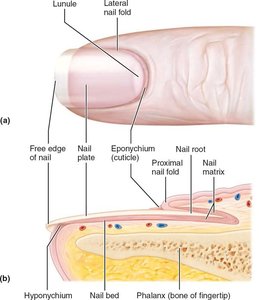
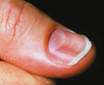
Nails are scale-like modifications of the epidermis containing hard keratin. They protect the distal phalanges and aid in sensation.
Root: Embedded in skin
Nail plate/body: Visible attached portion
Free edge: Extends beyond finger
Nail bed: Epidermis under nail plate
Nail matrix: Growth area
Nail folds: Skin overlapping nail border
Cuticle (eponychium): Projects onto nail body
Hyponychium: Under free edge, collects dirt
Skin Appendages: Glands
Sweat Glands
Eccrine (merocrine) sweat glands: Most numerous, thermoregulation, secrete watery sweat
Apocrine sweat glands: Axillary/anogenital areas, secrete viscous sweat, function as scent glands
Modified apocrine glands: Ceruminous (earwax), mammary (milk)
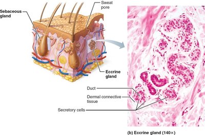
Oil (Sebaceous) Glands
Holocrine glands, secrete sebum into hair follicles
Lubricate skin and hair, bactericidal properties
Inactive until puberty, stimulated by androgens
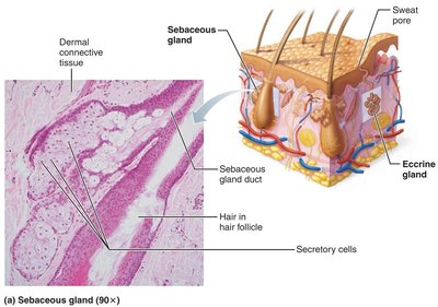
Summary Table: Cutaneous Glands
Gland Type | Function | Secretion | Method | Location |
|---|---|---|---|---|
Eccrine | Thermoregulation | Watery sweat | Merocrine | Palms, soles, forehead |
Apocrine | Scent | Viscous sweat | Merocrine | Axillary, anogenital |
Sebaceous | Lubrication, bactericidal | Sebum (oil) | Holocrine | Hair follicles, skin (except palms/soles) |
Functions of Skin
Protection
Chemical barriers: Sweat, sebum, acid mantle, melanin
Physical barriers: Keratinized cells, glycolipids
Biological barriers: Dendritic cells, macrophages
Body Temperature Regulation
Insensible perspiration: 500 ml/day
Sensible perspiration: Up to 12 L/day during heat
Vasoconstriction in cold, vasodilation in heat
Cutaneous Sensations
Touch: Meissner's corpuscles, Merkel cells
Pressure: Pacinian corpuscles
Hair movement: Hair follicle receptors
Pain: Free nerve endings
Metabolic Functions
Synthesizes vitamin D for calcium absorption
Disarms carcinogens, activates hormones
Makes collagenase for collagen turnover
Blood Reservoir
Holds up to 5% of blood volume
Can shunt blood to other organs
Excretion
Excretes nitrogenous wastes (ammonia, urea, uric acid)
Loss of salt and water via sweat
Skin Disorders: Cancer and Burns
Skin Cancer
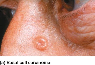
Basal cell carcinoma: Most common, least malignant, arises from stratum basale
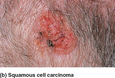
Squamous cell carcinoma: Second most common, can metastasize, arises from stratum spinosum
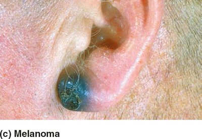
Melanoma: Cancer of melanocytes, most dangerous, highly metastatic
Burns
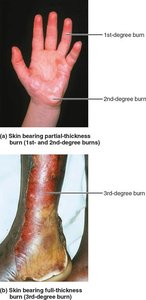
First-degree: Epidermal damage only, redness, pain
Second-degree: Epidermal and upper dermal damage, blisters
Third-degree: Entire skin thickness, gray-white, cherry red, or blackened, nerve endings destroyed
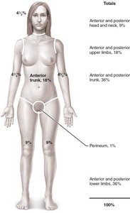
Critical if >25% second-degree, >10% third-degree, or face/hands/feet involved
Treatment: Debridement, antibiotics, temporary covering, skin grafts
Developmental Aspects
Infancy to Adulthood
Vernix caseosa: Protective substance in fetus
Milia: Small white spots in newborns
Skin thickens, gland activity increases, acne may develop
Optimal skin appearance in 20s–30s
Aging Skin
Epidermal replacement slows, skin thins, becomes dry and itchy
Subcutaneous fat and elasticity decrease, leading to wrinkles and cold intolerance
Increased risk of cancer due to decreased melanocytes and dendritic cells
Hair thinning
UV protection can delay aging
