 뒤로
뒤로The Integumentary System: Structure, Function, and Clinical Relevance
스터디 가이드 - 스마트 노트
 자료에 맞춘 맞춤형 노트, 핵심 정의, 예시, 맥락을 확장해 제공합니다.
The Integumentary System
Overview
The integumentary system is composed of the skin and its appendages, serving as the body's primary barrier against the external environment. It plays crucial roles in protection, sensation, thermoregulation, and metabolic functions.
Skin Layers: Epidermis, Dermis, Subcutaneous tissue
Appendages: Hair, nails, sweat glands, oil glands
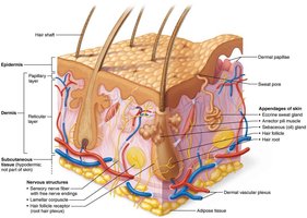
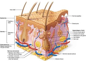
Skin Structure
Layers of the Skin
The skin consists of three main layers, each with distinct structural and functional properties.
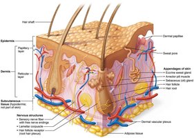
Epidermis: Outermost layer, composed of epithelial cells; provides a protective barrier.
Dermis: Middle layer, tough and leathery, mostly dense connective tissue; houses blood vessels, nerves, and glands.
Subcutaneous Tissue (Hypodermis): Deepest layer, primarily adipose tissue; insulates and anchors skin to underlying structures.
Cells of the Epidermis
Major Cell Types
The epidermis is a keratinized stratified squamous epithelium, avascular but innervated, containing four main cell types:
Keratinocytes: Produce keratin, the protein that gives skin its strength and waterproofing; tightly connected by desmosomes; continuously shed.
Melanocytes: Located in the deepest epidermis; produce melanin pigment, which protects keratinocyte DNA from UV damage.
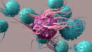
Dendritic (Langerhans) Cells: Star-shaped macrophages that patrol the epidermis; key activators of the immune system.
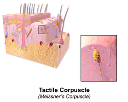
Tactile (Merkel) Cells: Sensory receptors for touch.
Layers of the Epidermis
Strata of the Epidermis
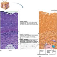
The epidermis is organized into distinct layers (strata), with thick skin (palms, soles) containing five layers and thin skin containing four.
Stratum Corneum: Most superficial; 20-30 layers of dead, keratinized cells.
Stratum Lucidum: Only in thick skin; few rows of flat, dead keratinocytes.
Stratum Granulosum: 1-5 layers of flattened cells; organelles deteriorate.
Stratum Spinosum: Several layers of keratinocytes unified by desmosomes.
Stratum Basale: Deepest layer; single row of actively mitotic stem cells.
Dermis
Structure and Function
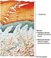
The dermis is a strong, flexible connective tissue layer containing fibroblasts, macrophages, mast cells, and white blood cells. It binds the body together and houses hair follicles, oil glands, and sweat glands.
Reticular Layer: Dense fibrous connective tissue; provides strength, elasticity, and hydration.
Papillary Dermis: Areolar connective tissue with interlacing collagen and elastic fibers; contains many small blood vessels.
Dermal Papillae
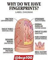
Dermal papillae are finger-like projections that interlock with the epidermis, forming ridges (fingerprints) that enhance grip.
Function: Increase surface area for exchange and provide friction for gripping.
Skin Color
Pigments and Clinical Significance
Skin color is determined by three main pigments:
Melanin: Produced by melanocytes; shields DNA from UV; more sun exposure increases melanin production.
Carotene: Yellow to orange pigment; accumulates in stratum corneum and hypodermis; can be converted to vitamin A.
Hemoglobin: Reddish pigment in blood; visible in fair skin due to transparency.
Clinical Signs
Changes in skin color can indicate disease:
Cyanosis: Blue skin due to low oxygenation of hemoglobin.
Pallor: Pale skin from anemia, low blood pressure, or emotional stress.
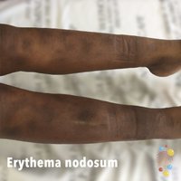
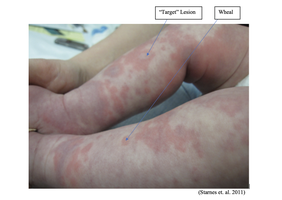
Erythema: Redness from fever, hypertension, inflammation, or allergy.
Jaundice: Yellow skin from liver disorders.
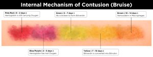
Bruises: Black-and-blue marks from clotted blood beneath skin; color changes as clot breaks down.

Skin Appendages: Cutaneous Glands
Sweat and Oil Glands
The skin contains two main types of sweat glands and oil glands, each with distinct structure and function.
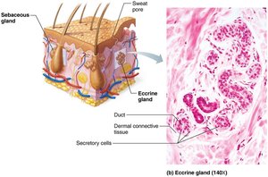
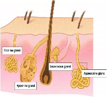
Eccrine (Merocrine) Sweat Glands: Most numerous; abundant on palms, soles, forehead; function in thermoregulation; secrete watery sweat (99% water, salts, vitamin C, antibodies, dermcidin, metabolic wastes).
Apocrine Sweat Glands: Confined to axillary and anogenital areas; secrete viscous, milky sweat with fatty substances and proteins; responsible for body odor; begin functioning at puberty.
Modified Apocrine Glands: Ceruminous glands (earwax), mammary glands (milk).
Sebaceous (Oil) Glands: Widely distributed except palms and soles; secrete sebum (oily, bactericidal, softens hair and skin); stimulated by hormones.
Functions of Skin
Protection
The skin acts as a barrier against microorganisms, abrasions, temperature extremes, and chemicals.
Chemical Barrier: Sweat, sebum, defensins, acid mantle, melanin.
Physical Barrier: Keratinized cells and glycolipids block water and solutes.
Biological Barrier: Phagocytic cells and DNA absorb UV radiation.
Body Temperature Regulation
Insensible Perspiration: 500 ml/day of unnoticeable sweat at rest.
Sensible Perspiration: Up to 12 L/day during overheating; cools the body.
Cold Response: Dermal blood vessels constrict to reduce heat loss.
Cutaneous Sensation
Sensory receptors in the skin detect touch, temperature, and pain.
Exteroreceptors: Respond to external stimuli.
Free Nerve Endings: Sense pain.
Metabolic Functions
Vitamin D Synthesis: Skin synthesizes vitamin D for calcium absorption.
Hormone Activation: Keratinocytes convert cortisone to hydrocortisone.
Collagenase Production: Aids in collagen turnover, preventing wrinkles.
Blood Reservoir
Skin can hold up to 5% of total blood volume; vessels constrict to shunt blood to other organs.
Excretion
Skin excretes nitrogenous wastes (ammonia, urea, uric acid) and loses salt and water through sweat.
Clinical Relevance: Skin Cancer and Burns
Skin Cancer
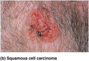
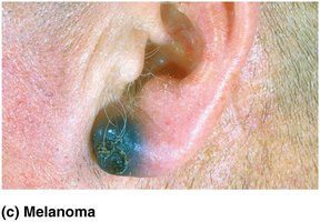
Skin cancer is a major concern, with three primary types:
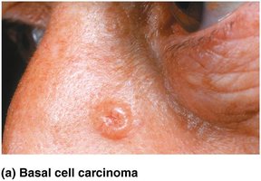
Basal Cell Carcinoma: Least malignant, most common; arises from stratum basale; cured by excision.
Squamous Cell Carcinoma: Second most common; can metastasize; arises from stratum spinosum; treated by surgery or radiation.
Melanoma: Most dangerous; cancer of melanocytes; highly metastatic and resistant to chemotherapy; treated by excision and immunotherapy.
Burns
Burns are classified by severity and can be life-threatening due to dehydration and electrolyte imbalance.
First-degree: Epidermal damage only; redness, edema, pain.
Second-degree: Epidermal and upper dermal damage; blisters.
Third-degree: Entire thickness of skin; gray-white, cherry red, or blackened; no pain (nerve endings destroyed); skin grafting required.
Critical Burns: >25% body with second-degree, >10% with third-degree, or third-degree on face, hands, feet.
Treatment: Debridement, antibiotics, temporary covering, skin grafts.
Summary Table: Layers and Functions of the Skin
Layer | Main Tissue Type | Key Functions |
|---|---|---|
Epidermis | Stratified squamous epithelium | Protection, waterproofing, UV defense |
Dermis (Papillary) | Areolar connective tissue | Support, blood supply, sensory input |
Dermis (Reticular) | Dense irregular connective tissue | Strength, elasticity, hydration |
Subcutaneous (Hypodermis) | Adipose tissue | Insulation, energy storage, anchoring |
Summary Table: Skin Glands
Gland Type | Location | Secretion | Function |
|---|---|---|---|
Eccrine Sweat | Most skin areas | Watery sweat | Thermoregulation |
Apocrine Sweat | Axillary, anogenital | Milky, viscous sweat | Body odor |
Sebaceous (Oil) | Hair follicles | Sebum | Lubrication, antibacterial |
Ceruminous | Ear canal | Cerumen (earwax) | Protection |
Mammary | Breasts | Milk | Nourishment |
Key Equations
Surface Area Calculation (for burns)
The Rule of Nines is used to estimate the percentage of body surface area affected by burns: Example: Anterior trunk (18%), each arm (9%), each leg (18%), head (9%), perineum (1%).
Conclusion
The integumentary system is essential for protection, homeostasis, and clinical diagnosis. Understanding its structure and function is fundamental for recognizing disease states and managing skin-related conditions.
