 뒤로
The Lymphatic System and Immune Systems: Structure, Function, and Defense
스터디 가이드 - 스마트 노트
 자료에 맞춘 맞춤형 노트, 핵심 정의, 예시, 맥락을 확장해 제공합니다.
The Lymphatic System
Overview and Structure
The lymphatic system is a network of vessels and organs that plays a crucial role in fluid balance, immune defense, and fat absorption. It works closely with the cardiovascular system to return excess interstitial fluid to the bloodstream and to filter pathogens.
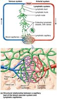
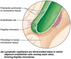
Lymphatic vessels are one-way, blind-ended capillaries that absorb fluid left behind in tissues.
Interstitial fluid entering a lymphatic vessel is called lymph.
Endothelial cells overlap to form mini-valves that regulate fluid entry.
Collagen filaments anchor cells to tissue, preventing collapse.
Lymphatic capillaries progress to collecting vessels, trunks, and finally ducts.
Lymphatic Drainage
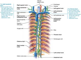
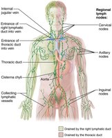
Lymphatic drainage is organized into two main ducts:
Right lymphatic duct: Drains the right arm, right side of the head, and thorax. Joins circulation at the junction of the right internal jugular and subclavian veins.
Thoracic duct: Drains lymph from the rest of the body. Begins as the cisterna chyli (a sac anterior to L1-L2) and joins circulation at the junction of the left internal jugular and subclavian veins.
Functions of the Lymphatic System
The lymphatic system serves several essential functions:
Returns excess tissue fluid and leaked proteins to the bloodstream.
Collects and transports pathogens to lymph nodes for immune surveillance.
Carries absorbed fat from the small intestine to the thoracic duct via lacteals.
Lymph Transport
Lymph vessels operate under low pressure and rely on several mechanisms for transport:
Skeletal muscle pump
Respiratory pump
Valves in collecting vessels prevent backflow
Trunks and ducts contain smooth muscle for propulsion
Lymphoid Tissues and Organs
Lymphoid tissues and organs are composed of reticular connective tissue and immune cells.
Lymph nodes: Clustered along vessels, act as lymph filters, and activate the immune system.
Contain B cells, T cells, macrophages, and dendritic cells in cortex and medulla.
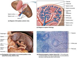
The Spleen
The spleen is the largest lymphoid organ and has four main functions:
White pulp: Lymphocyte proliferation and immune surveillance.
Red pulp: Macrophages extract aged blood cells and pathogens.
Recycles erythrocytes; stores iron, bilirubin, and amino acids.
Stores platelets and monocytes.
The Thymus
The thymus is located deep to the sternum and is most active during childhood. It secretes hormones (thymopoietin, thymulin, thymosins) essential for T cell maturation.
Mucosa-Associated Lymphoid Tissue (MALT)
MALT protects open cavities and includes:
Tonsils
Peyer's patches
Bronchi
Genitourinary tract
Appendix
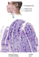
Tonsils
Tonsils are not fully encapsulated and are located at strategic points in the pharynx and oral cavity.
Epithelium forms crypts that trap and destroy bacteria, enhancing immunity.
Peyer's Patches
Peyer's patches are clusters of lymphoid follicles in the distal small intestine, important for immune defense and pathogen destruction.
The Immune System
Innate Immunity
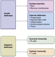
Innate immunity is present at birth and provides immediate, nonspecific defense.
First line: Surface barriers (skin, mucosa) and their secretions.
Second line: Inflammation, phagocytes, leukocytosis, and fever.
Inflammation
Inflammation is a local, nonspecific response to tissue damage or infection.
Chemical mediators (histamine, kinins, complement, prostaglandins, leukotrienes) trigger vasodilation and vasopermeability.
Cardinal signs: Heat, redness, pain, swelling.
Mobilization of Phagocytes
Leukocytosis: Increased WBCs in blood.
Margination: Neutrophils cling to vessel walls.
Diapedesis: Neutrophils squeeze out of capillaries.
Chemotaxis: Neutrophils follow chemical signals to injury site.
Phagocytosis
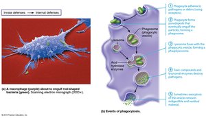
Phagocytes (neutrophils, macrophages) engulf pathogens, aided by antibodies or complement.
Pseudopods form a phagosome.
Lysosomes fuse to form a phagolysosome, releasing enzymes and oxidizing chemicals.
Pathogen is destroyed and residual material is exocytosed.
Systemic Response
Leukocytosis, lymphadenopathy, and fever (pyrogens reset hypothalamic set point).
Fever enhances lymphocyte activity, increases metabolism, and inhibits microbes.
Adaptive Immunity
Adaptive immunity is a specific, systemic response with memory, beginning 48-72 hours after infection.
Produces antibodies to specific antigens.
Generates T and B memory cells.
Antibodies circulate systemically.
Branches of Adaptive Immunity
Humoral immunity: B cells differentiate into plasma cells, releasing antibodies targeting extracellular antigens.
Cell-mediated immunity: T cells target infected or abnormal cells.
Antigen and Lymphocyte Development
An antigen is any substance provoking an immune response; contains multiple antigenic determinants (epitopes).
T and B lymphocytes originate from red bone marrow; B cells mature in bone marrow, T cells in thymus.
Lymphocytes develop immunocompetence and self-tolerance, residing in lymph nodes and organs.
Major Histocompatibility Complex (MHC)
MHC I: On all cells except erythrocytes.
MHC II: On antigen-presenting cells (APCs).
T cells must recognize MHC receptors and not self-antigens to avoid apoptosis.
Antigen Encounter and Presentation
T cells circulate and migrate to tissues.
Lymphatic system delivers antigens to lymph nodes and organs.
APCs (dendritic cells, macrophages) phagocytose antigens, display fragments on MHC, and present to T cells.
Primary and Secondary Humoral Immunity
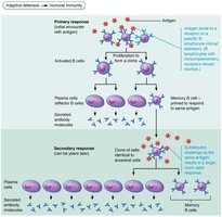
Primary response: B cells encounter antigen, proliferate, and differentiate into plasma cells and memory B cells. Plasma cells produce antibodies (immunoglobulins).
Secondary response: Memory B cells respond rapidly to the same antigen, producing more antibody quickly.
Immunological Memory
Primary response: Begins in 3-6 days, IgM produced first, IgG more abundant.
Secondary response: Faster (1-3 days), larger, more IgG than IgM.
Antibody Structure and Classes
Each antibody monomer has two antigen binding sites; stem region can be embedded in cell membranes.
Five classes: IgM (first, agglutination), IgA (secretions), IgD (B cell receptor), IgG (most abundant, crosses placenta), IgE (allergic reactions).
Antibody Functions
Antibodies inactivate pathogens by:
Neutralization: Masking dangerous parts of toxins or viruses.
Agglutination: Clumping antigens together.
Precipitation: Forming insoluble complexes.
All enhance phagocytosis (opsonization).
Cell-Mediated Immunity
T lymphocytes target cells with intracellular pathogens or abnormal cells.
Helper T cells (TH): Mobilize B cells and cytotoxic T cells; have CD4 receptors recognizing MHC II.
Cytotoxic T cells (TC): Destroy infected/cancerous cells; have CD8 receptors recognizing MHC I.
Regulatory T cells: Suppress immune responses.
Activation and Response
TH2 cells stimulate strong humoral response by binding antigen on B cell MHC II, releasing cytokines for B cell activation.
Cytotoxic T cells release perforins and granzymes to induce apoptosis in infected cells.
Immunity Type | Key Cells | Target |
|---|---|---|
Innate | Phagocytes, NK cells | General pathogens |
Humoral (Adaptive) | B cells, plasma cells | Extracellular antigens |
Cell-mediated (Adaptive) | T cells | Infected/abnormal cells |
Summary
The lymphatic and immune systems are essential for fluid balance, pathogen defense, and immune memory. Their coordinated actions protect the body from infection and disease, and their structure and function are fundamental to understanding human physiology.
