 뒤로
뒤로The Lymphatic System and Lymphoid Organs: Structure, Function, and Clinical Relevance
스터디 가이드 - 스마트 노트
 자료에 맞춘 맞춤형 노트, 핵심 정의, 예시, 맥락을 확장해 제공합니다.
The Lymphatic System: Overview
Functions of the Lymphatic System
The lymphatic system is essential for maintaining fluid balance, immune defense, and the absorption of dietary fats. It returns interstitial fluid and leaked plasma proteins back to the blood, provides structural support for immune cells, and houses lymphoid organs and tissues.
Fluid Recovery: Returns excess interstitial fluid (~3L/day) to the bloodstream.
Immune Response: Provides sites for immune cell activation and proliferation.
Absorption of Fats: Specialized lymphatic capillaries (lacteals) absorb dietary fats in the intestine.
Components of the Lymphatic System
Lymphatic vessels: Network of drainage vessels transporting lymph.
Lymph: Fluid within lymphatic vessels.
Lymph nodes: Filter lymph and house immune cells.
Lymphoid organs: Include spleen, thymus, tonsils, and other tissues.
Structure and Distribution of Lymphatic Vessels
Lymphatic Capillaries
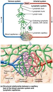
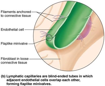
Lymphatic capillaries are blind-ended tubes interwoven with blood capillaries and tissue cells. They are highly permeable, allowing uptake of large molecules and particles, including proteins, cell debris, pathogens, and cancer cells.
Minivalves: Overlapping endothelial cells form one-way valves, opening with increased extracellular fluid (ECF) volume.
Anchoring Filaments: Collagen filaments anchor capillaries to connective tissue, facilitating valve function.
Lacteals: Specialized capillaries in intestinal mucosa absorb digested fats.
Larger Lymphatic Vessels
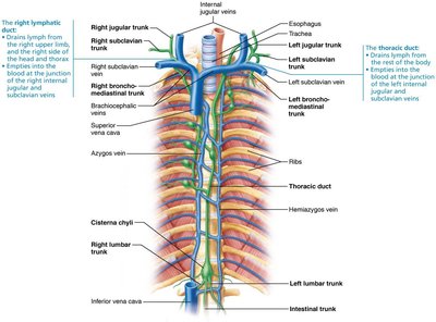
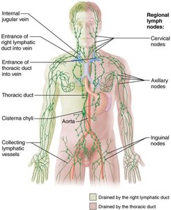
Lymphatic capillaries drain into larger collecting vessels, trunks, and ducts. These vessels have thinner walls and more internal valves than veins, and they anastomose more frequently.
Collecting Vessels: Travel with superficial veins or deep arteries.
Lymphatic Trunks: Drain large regions of the body (lumbar, bronchomediastinal, subclavian, jugular, intestinal).
Lymphatic Ducts:
Right lymphatic duct: Drains right upper limb, right side of head and thorax.
Thoracic duct: Drains rest of the body, often begins as cisterna chyli.
Venous Return: Ducts empty lymph into venous circulation at the junction of internal jugular and subclavian veins.
Source and Transport of Lymph
Mechanisms of Lymph Transport
Lymph transport is a low-pressure system, similar to veins. Lymph is propelled by several mechanisms:
Skeletal muscle contraction (milking action)
Respiratory movements: Pressure changes during breathing
Valves: Prevent backflow
Arterial pulsations: Nearby arteries help propel lymph
Smooth muscle contraction: In vessel walls
Physical activity increases lymph flow, while immobilization slows it, aiding local healing.
Lymphoid Cells and Tissue
Lymphoid Cells
Lymphoid cells are divided into immune system cells and supporting cells.
Lymphocytes: Adaptive immune cells; mature into T cells (manage immune response, attack infected cells) and B cells (produce antibodies).
Macrophages: Phagocytize foreign substances, activate T cells.
Dendritic cells: Capture antigens, deliver to lymph nodes, activate T cells.
Reticular cells: Produce reticular fibers (stroma), providing structural support.
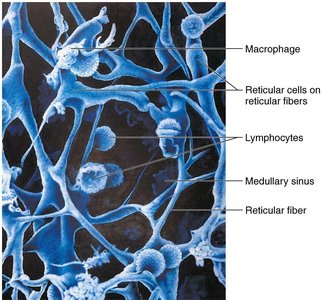
Lymphoid Tissue
Lymphoid tissue houses and provides proliferation sites for lymphocytes, offering surveillance points for immune cells. It is largely composed of reticular connective tissue.
Lymphoid follicles (nodules): Spherical bodies with germinal centers of proliferating B cells.
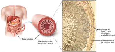
Peyer's patches and appendix: Isolated aggregations of lymphoid tissue.
Lymphoid Organs
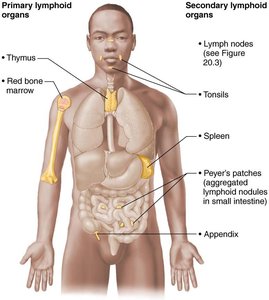
Primary vs. Secondary Lymphoid Organs
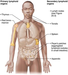
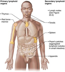
Lymphoid organs are classified as primary (sites of lymphocyte maturation) or secondary (sites of lymphocyte activation).
Primary: Red bone marrow (B cell maturation), thymus (T cell maturation)
Secondary: Lymph nodes, spleen, MALT, tonsils, Peyer's patches, appendix
Lymph Nodes
Structure and Function
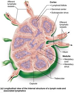
Lymph nodes are bean-shaped, less than 2.5 cm, surrounded by a fibrous capsule. They filter lymph and provide sites for immune activation.
Cleansing: Macrophages remove microorganisms and debris.
Immune Activation: Lymphocytes mount attacks against antigens.
Histological Regions
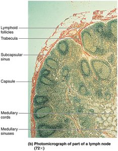
Cortex: Contains follicles with germinal centers (B cells), deep cortex houses T cells and dendritic cells.
Medulla: Medullary cords with B cells, T cells, plasma cells; lymph sinuses with macrophages.
Circulation in Lymph Nodes
Lymph enters via afferent vessels, passes through sinuses, exits via efferent vessels at the hilum.
Fewer efferent vessels cause stagnation, allowing immune cells time to act.
Spleen
Structure and Function
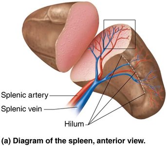
The spleen is the largest lymphoid organ, located in the left abdominal cavity. It is blood-rich and serves multiple functions:
Immune Surveillance: Site for lymphocyte proliferation and response.
Blood Cleansing: Removes aged blood cells and platelets; macrophages remove debris.
Storage: Stores breakdown products of RBCs, platelets, and monocytes.
Fetal Function: May produce erythrocytes in fetus.
Histological Components
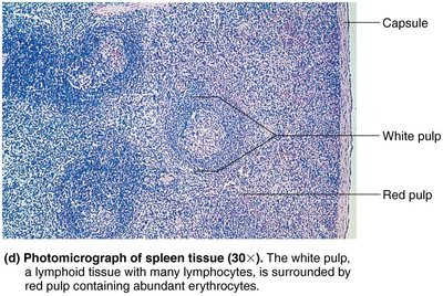
White pulp: Immune function, clusters of lymphocytes around central arteries.
Red pulp: Destruction of old blood cells and pathogens, rich in RBCs and macrophages.
MALT (Mucosa-Associated Lymphoid Tissue)
Structure and Function
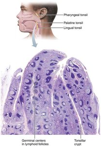
MALT consists of lymphoid tissues in mucous membranes throughout the body, protecting against pathogens entering via respiratory, genitourinary, and digestive tracts.
Tonsils: Form ring around pharynx, gather and remove pathogens in food/air.
Peyer's patches: Clusters in distal small intestine, destroy bacteria, generate memory lymphocytes.
Appendix: Offshoot of large intestine, similar function to Peyer's patches.
Thymus
Structure and Function
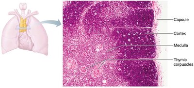
The thymus is a bilobed organ in the inferior neck and mediastinum, most active in childhood. It is the site of T cell maturation and differs from other lymphoid organs:
No follicles: Lacks B cells.
Does not directly fight antigens: Functions strictly in T cell maturation.
Stroma: Made of epithelial cells, not reticular fibers.
Blood-thymus barrier: Isolates immature T cells from antigens.
Summary Table: Lymphoid Organs and Tissues
The following table summarizes the major functions, histological features, and special characteristics of lymphoid organs and tissues:
Organ | Major Functions | Capsule | Cortex & Medulla | Lymphoid Follicles | Stroma | Special Features |
|---|---|---|---|---|---|---|
Lymph nodes | Cleanses lymph; site for lymphocyte activation and proliferation | Yes | Yes | Yes (in cortex) | Reticular connective tissue | Both afferent and efferent lymphatic vessels |
Spleen | Cleanses blood; removes aged/defective RBCs; stores platelets, monocytes; immune activation | Yes | Yes | Yes (in white pulp) | Reticular connective tissue | Red and white pulp |
MALT* | Prevents pathogens from penetrating mucosa; site for lymphocyte activation and proliferation | No | No | Yes | Reticular connective tissue | Diffuse lymphoid tissue |
Thymus | Site of T cell maturation | Yes | Yes | No | Epithelial tissue | Thymic corpuscles |
*Red bone marrow, a primary lymphoid organ, is not included. MALT = mucosa-associated lymphoid tissues.
Key Terms and Concepts
Lymph: Fluid transported by lymphatic vessels, derived from interstitial fluid.
Antigen: Any substance recognized as foreign by the immune system.
Stroma: Structural network supporting immune cells in lymphoid organs.
Germinal center: Area of proliferating B cells within lymphoid follicles.
Minivalve: One-way valve formed by overlapping endothelial cells in lymphatic capillaries.
Equations and Formulas
While the lymphatic system does not rely heavily on mathematical equations, fluid dynamics principles apply:
Fluid balance equation:
Volume of lymph returned:
Clinical Relevance
Lymphedema: Swelling due to lymphatic obstruction.
Metastasis: Cancer cells may travel via lymphatic vessels.
Immunodeficiency: Disorders affecting lymphoid organs can impair immune function.
Summary
The lymphatic system is vital for fluid balance, immune defense, and fat absorption. Its vessels, nodes, and organs work together to filter lymph, activate immune responses, and protect the body from pathogens.
