 뒤로
뒤로The Muscular System: Anatomy, Histology, and Physiology
스터디 가이드 - 스마트 노트
 자료에 맞춘 맞춤형 노트, 핵심 정의, 예시, 맥락을 확장해 제공합니다.
The Muscular System
Overview of Muscle Functions
The muscular system is essential for movement, posture, joint stabilization, heat production, control of body openings, movement of substances, and protection/support of organs. Muscles perform both voluntary and involuntary actions, contributing to overall body function.
Movement: Muscles contract to move bones and body parts.
Posture: Muscles maintain body position.
Joint Stabilization: Muscles reinforce joints.
Heat Production: Muscle activity generates heat.
Control of Openings: Sphincter muscles regulate passageways.
Movement of Substances: Muscles move blood, food, and other substances.
Protection/Support: Muscles shield internal organs.
Universal Characteristics of Muscle
Muscle tissue possesses five universal characteristics that enable its function:
Excitability: Ability to respond to stimuli.
Conductivity: Ability to transmit electrical signals.
Contractility: Ability to shorten and generate force.
Extensibility: Ability to stretch without damage.
Elasticity: Ability to return to original shape after stretching.
Types of Muscle Tissue
Classification and Comparison
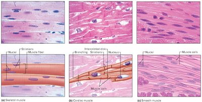
There are three types of muscle tissue in the human body: skeletal, cardiac, and smooth muscle. Each type has distinct anatomical and functional properties.
Skeletal Muscle: Voluntary, striated, multinucleated, attached to bones.
Cardiac Muscle: Involuntary, striated, single nucleus, found only in the heart.
Smooth Muscle: Involuntary, non-striated, single nucleus, found in walls of hollow organs.
Key Terms: myocyte (muscle cell), muscle fiber (skeletal/smooth), cardiomyocyte (cardiac).
Type | Shape | Striations | Control | Function |
|---|---|---|---|---|
Skeletal | Long | Yes | Voluntary | Locomotion, heat |
Cardiac | Short, branched | Yes | Involuntary | Pumping blood |
Smooth | Fusiform | No | Involuntary | Peristalsis, control of openings |
Muscle Anatomy: Shapes and Structure
Shapes of Whole Muscles
Muscles are classified by their overall shape, which influences their function and location in the body.
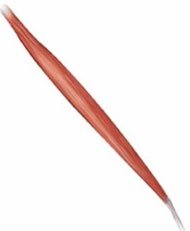
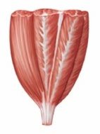
Fusiform: Spindle-shaped, thick in the middle and tapered at ends.
Parallel: Uniform width, parallel fascicles.
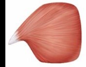
Convergent: Broad origin, fascicles converge toward a single tendon.
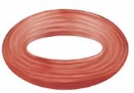
Circular: Fascicles arranged in concentric rings (sphincters).
Pennate: Fascicles attach obliquely to a tendon (unipennate, bipennate, multipennate).
Muscle Vocabulary
Prime mover (agonist): Main muscle responsible for an action.
Synergist: Helper muscle for a given action.
Antagonist: Muscle that opposes the action.
Fixator: Muscle that stabilizes a bone for the prime mover.
Innervation
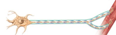
Innervation refers to the nerve supply to a muscle, which is essential for muscle contraction. Motor neurons transmit signals to muscle fibers, causing them to contract.
Muscle Histology and Microanatomy
Gross Structure of Skeletal Muscle
Skeletal muscle is organized hierarchically:
Whole Muscle → Fascicle → Muscle Fiber (Cell) → Myofibril → Myofilament
Connective tissue investments include:
Epimysium: Surrounds entire muscle.
Perimysium: Wraps fascicles.
Endomysium: Surrounds individual muscle fibers.
Microscopic Structure of Muscle Tissue
Muscle tissue can be distinguished under the microscope by cell shape, striations, and nuclei arrangement.
Skeletal muscle: Long, multinucleated, striated fibers.
Cardiac muscle: Short, branched, striated, single nucleus, intercalated discs.
Smooth muscle: Spindle-shaped, non-striated, single nucleus.
Muscle Physiology
Excitation-Contraction Coupling
Muscle contraction is initiated by electrical signals from motor neurons. The process involves:
Excitation: Motor neuron releases acetylcholine (ACh) at the neuromuscular junction, depolarizing the sarcolemma.
Excitation-Contraction Coupling: Action potential travels along T-tubules, triggering calcium release from the sarcoplasmic reticulum.
Contraction: Calcium binds to troponin, moving tropomyosin and exposing actin binding sites. Myosin heads bind actin, powered by ATP, causing sarcomere shortening (sliding filament theory).
Relaxation: Nerve stimulation ends, ACh is broken down, calcium is reabsorbed, and muscle returns to resting state.
Sliding Filament Theory
The sliding filament theory explains how sarcomeres contract:
Actin filaments slide over myosin filaments toward the M-line.
Requires ATP and Ca2+.
Thick and thin filaments do not change length; the sarcomere shortens.
Cross-Bridge Cycle
The cross-bridge cycle is the sequence of events during muscle contraction:
Myosin binds to actin (cross-bridge formation).
Powerstroke: Myosin head pivots, pulling actin toward the M-line.
Myosin releases from actin when new ATP binds.
Myosin recharges by hydrolyzing ATP to ADP + Pi.
Energy for Muscle Contraction
Muscle fibers use ATP for contraction, which can be generated by:
Creatine Phosphate: Provides rapid, short-term energy.
Anaerobic Cellular Respiration: Produces ATP without oxygen (glycolysis, lactic acid formation).
Aerobic Cellular Respiration: Produces ATP with oxygen in mitochondria, yielding more ATP for sustained activity.
Clinical Applications
Rigor Mortis
Rigor mortis is the postmortem stiffening of muscles due to calcium leakage and lack of ATP, preventing muscle relaxation.
Botox
Botox prevents muscle contraction by blocking acetylcholine release at the neuromuscular junction, causing flaccid paralysis.
Summary Table: Muscle Tissue Types
Type | Shape | Striations | Control | Function |
|---|---|---|---|---|
Skeletal | Long | Yes | Voluntary | Locomotion, heat |
Cardiac | Short, branched | Yes | Involuntary | Pumping blood |
Smooth | Fusiform | No | Involuntary | Peristalsis, control of openings |
Key Equations
Sodium/Potassium Pump:
ATP Hydrolysis:
Muscle Fiber Resting Membrane Potential:
Additional info:
Some content was inferred and expanded for clarity and completeness, including detailed explanations of muscle contraction stages, clinical applications, and energy sources for muscle contraction.
