 뒤로
The Muscular System: Structure, Function, and Physiology
스터디 가이드 - 스마트 노트
 자료에 맞춘 맞춤형 노트, 핵심 정의, 예시, 맥락을 확장해 제공합니다.
The Muscular System
Introduction to Muscle Tissue
Muscle tissue is one of the four primary tissue types in the human body, specialized for contraction and responsible for movement. There are three types of muscle tissue: skeletal, cardiac, and smooth muscle. This chapter focuses on the structure, function, and physiology of skeletal muscle, with comparisons to cardiac and smooth muscle where relevant.
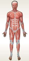
Functions of Skeletal Muscle Tissue
Movement of the Skeleton: Skeletal muscles pull on tendons to move bones, enabling voluntary movement.
Posture and Body Position: Continuous muscle contractions maintain posture.
Support of Soft Tissues: Muscles form the walls of the abdominal and pelvic cavities, supporting internal organs.
Guarding Entrances and Exits: Muscles encircle openings of the digestive and urinary tracts, controlling passage of materials.
Maintenance of Body Temperature: Muscle contractions generate heat, helping to maintain body temperature.
Organization of Skeletal Muscle Tissue
Structural Organization
Skeletal muscles are complex organs composed of muscle tissue, connective tissues, blood vessels, and nerves. Each skeletal muscle cell is called a muscle fiber.
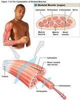
Connective Tissue Layers
Epimysium: Surrounds the entire muscle.
Perimysium: Divides the muscle into fascicles (bundles of muscle fibers) and contains blood vessels and nerves.
Endomysium: Covers each muscle fiber and ties fibers together; contains capillaries and nerve fibers.
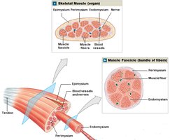
Connective Tissue Attachments
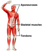
Tendon: A bundle of collagen fibers attaching muscle to bone.
Aponeurosis: A broad sheet of collagen fibers connecting muscles to each other.
Blood Vessels and Nerves
Skeletal muscles have an extensive network of blood vessels to meet high energy demands.
They are under voluntary control, requiring stimulation by the central nervous system.
Axons (nerve fibers) innervate individual muscle fibers.
Microscopic Anatomy of Skeletal Muscle Fibers
Features of Skeletal Muscle Fibers
Muscle fibers can be very long (up to 60 cm) and are multinucleate.
They have a striated appearance due to the arrangement of myofilaments.
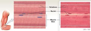
Sarcolemma and Transverse Tubules (T Tubules)
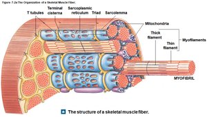
Sarcolemma: The plasma membrane of a muscle fiber, surrounding the sarcoplasm (cytoplasm).
T Tubules: Invaginations of the sarcolemma that transmit electrical impulses deep into the fiber, ensuring coordinated contraction.
Myofibrils and Myofilaments
Myofibrils: Cylindrical structures running the length of the muscle fiber, composed of bundles of thick and thin myofilaments.
The sliding of these filaments causes muscle contraction.
Sarcoplasmic Reticulum (SR) and Triads
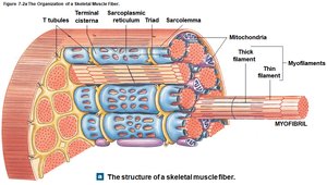
Sarcoplasmic Reticulum (SR): Specialized smooth endoplasmic reticulum that stores calcium ions.
Terminal Cisternae: Expanded regions of the SR adjacent to T tubules.
Triad: A structure formed by a T tubule and two terminal cisternae, crucial for excitation-contraction coupling.
Sarcomeres: The Functional Unit
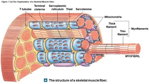
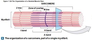
The sarcomere is the smallest functional unit of a muscle fiber, responsible for contraction. Each myofibril contains thousands of sarcomeres arranged end to end.
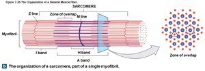
Sarcomere Lines and Bands
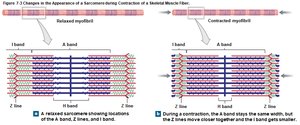
Z lines: Define the boundaries of each sarcomere; connected by titin to thick filaments.
M line: Center of the sarcomere, connecting thick filaments.
A band: Dark region containing thick filaments (with some overlap of thin filaments).
I band: Light region containing only thin filaments, includes the Z line.
H band: Central region with only thick filaments, includes the M line.
Myofilament Structure
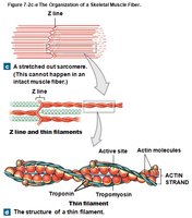
Thin Filaments
Composed mainly of actin molecules, with specific active sites for myosin binding.
At rest, active sites are covered by tropomyosin, which is held in place by troponin.
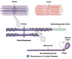
Thick Filaments
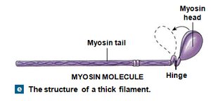
Composed of myosin molecules, each with a tail and a globular head.
Myosin heads interact with actin during contraction, forming cross-bridges.
Mechanism of Muscle Contraction
Sliding Filament Theory
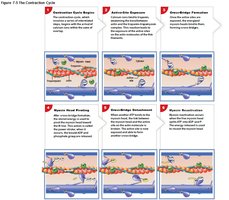
Muscle contraction occurs as thin filaments slide toward the center of the sarcomere, while thick filaments remain stationary. This shortens the sarcomere and generates tension.
Cross-Bridge Cycle
Myosin heads bind to actin, forming cross-bridges.
Heads pivot, pulling thin filaments toward the center of the sarcomere.
Cross-bridges detach and reset, repeating the cycle as long as ATP and calcium are available.
The Neuromuscular Junction (NMJ)
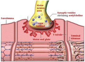
The NMJ is the synapse between a motor neuron and a skeletal muscle fiber. It consists of the axon terminal (containing acetylcholine, ACh), the synaptic cleft, and the motor end plate of the muscle fiber.
Steps of Muscle Contraction and Relaxation
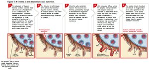
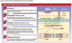
ACh is released from the neuron, triggering an action potential in the sarcolemma.
The action potential travels down T tubules, causing the SR to release Ca2+.
Ca2+ binds to troponin, shifting tropomyosin and exposing actin active sites.
Myosin heads bind to actin, initiating the contraction cycle.
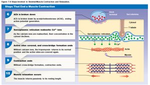
Relaxation occurs when ACh is broken down, Ca2+ is reabsorbed, and active sites are covered again.
Muscle Tension and Contraction Types
Muscle Twitch and Myogram
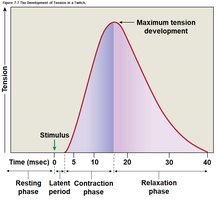
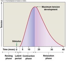
A muscle twitch is a single stimulus-contraction-relaxation cycle in a muscle fiber. The myogram is a graph showing tension development during a twitch.
Summation and Tetanus
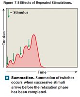
Summation: Addition of one twitch to another, resulting in greater tension.
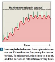
Incomplete Tetanus: Rapid cycles of contraction and relaxation, producing almost peak tension.
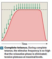
Complete Tetanus: No relaxation phase; maximum tension is produced.
Motor Units and Recruitment
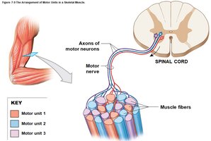
Motor Unit: A single motor neuron and all the muscle fibers it controls.
Recruitment is the activation of additional motor units to increase tension.
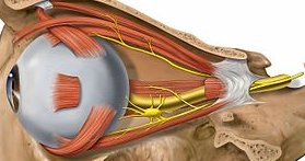
Small motor units allow precise control; large units produce powerful contractions.
Types of Muscle Contraction
Isotonic Contraction: Muscle changes length (e.g., lifting a book).
Isometric Contraction: Muscle length remains the same (e.g., pushing against a wall).
Muscle Tone and Atrophy
Muscle Tone: Resting tension that stabilizes bones and joints.
Atrophy: Reduction in muscle size and strength due to lack of stimulation.
Muscle Metabolism and Energy
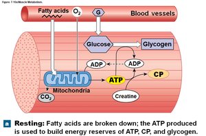
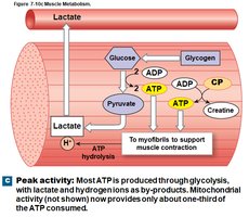
ATP and Creatine Phosphate
Muscle contraction requires ATP.
Excess ATP is stored as creatine phosphate (CP) for rapid regeneration of ATP during contraction.
Aerobic and Anaerobic Metabolism
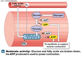
Aerobic Metabolism: Requires oxygen, occurs in mitochondria, and produces most ATP during rest and moderate activity.
Anaerobic Glycolysis: Occurs in the cytoplasm without oxygen, produces less ATP, and leads to lactic acid buildup.
Muscle Fatigue and Recovery
Muscle Fatigue: Inability to contract due to energy depletion or pH changes (lactic acid buildup).
Recovery Period: Time required to restore normal conditions, including oxygen debt repayment and lactic acid removal.
Muscle Fiber Types and Performance
Fast-Twitch vs. Slow-Twitch Fibers
Fast-Twitch (Type II): Large diameter, rapid contraction, fatigue quickly, rely on glycolysis.
Slow-Twitch (Type I): Smaller diameter, slower contraction, fatigue resistant, rely on aerobic metabolism, rich in myoglobin.
Muscle color varies: white muscles (fast fibers), red muscles (slow fibers), most human muscles are mixed (pink).
Muscle Conditioning
Anaerobic Endurance: Increased by brief, intense activity (e.g., weightlifting).
Aerobic Endurance: Increased by sustained, low-intensity activity (e.g., running).
Types of Muscle Tissue
Cardiac Muscle Tissue
Found only in the heart; cells are striated, branched, and connected by intercalated discs with gap junctions.
Exhibits automaticity (contracts without neural input), longer contractions, and cannot undergo tetanus.
Relies on aerobic metabolism and extracellular calcium for contraction.
Smooth Muscle Tissue
Found in walls of most organs; cells are spindle-shaped, non-striated, and have a single nucleus.
Lacks sarcomeres; contraction causes twisting motion.
Can contract automatically or in response to neural/hormonal signals.
Comparison Table: Skeletal, Cardiac, and Smooth Muscle
Feature | Skeletal Muscle | Cardiac Muscle | Smooth Muscle |
|---|---|---|---|
Location | Attached to bones | Heart | Walls of organs |
Striations | Yes | Yes | No |
Nuclei | Multinucleate | Single (usually) | Single |
Control | Voluntary | Involuntary | Involuntary |
Contraction | Rapid, short | Moderate, rhythmic | Slow, sustained |
Muscle Naming and Actions
Muscle Origins, Insertions, and Actions
Origin: Fixed attachment point of the muscle.
Insertion: Movable attachment point.
Action: The movement produced (e.g., flexion, extension).
Muscle Roles
Prime Mover (Agonist): Main muscle responsible for movement.
Antagonist: Opposes the prime mover.
Synergist: Assists the prime mover.
Fixator: Stabilizes the origin of the prime mover.
Basis for Muscle Names
Names may indicate location, shape, size, direction of fibers, number of origins, or action (e.g., flexor carpi radialis longus).
Major Muscles of the Body
Axial Muscles
Include muscles of the head, neck, spine, trunk, and pelvic floor.
Examples: orbicularis oris (mouth), temporalis (chewing), erector spinae (spine extension), diaphragm (breathing).
Appendicular Muscles
Muscles of the shoulder, upper and lower limbs, and pelvic girdle.
Examples: deltoid (arm abduction), biceps brachii (elbow flexion), quadriceps femoris (knee extension), gastrocnemius (plantar flexion).
Effects of Aging and System Integration
Aging Effects on Muscle
Muscle fibers decrease in size and elasticity.
Increased fibrous tissue, decreased exercise tolerance, and slower recovery from injury.
Muscle System Integration
Muscular, cardiovascular, respiratory, integumentary, nervous, and endocrine systems work together during exercise to maintain homeostasis.
