 뒤로
The Muscular System: Structure, Function, and Classification
스터디 가이드 - 스마트 노트
 자료에 맞춘 맞춤형 노트, 핵심 정의, 예시, 맥락을 확장해 제공합니다.
The Muscular System
Introduction
The muscular system is a fundamental component of human anatomy, responsible for movement, posture, and various physiological functions. Skeletal muscles are the most prominent type, enabling voluntary movements and contributing to homeostasis.
Structure of Skeletal Muscle
Muscle Organization and Connective Tissue
Skeletal muscle is composed of muscle fibers, blood vessels, nerves, and connective tissue. The organization of these components is hierarchical, providing both structural integrity and functional specialization.
Muscle cell (fiber): The basic unit, surrounded by endomysium.
Fascicle: Bundle of muscle fibers, surrounded by perimysium.
Muscle: All fascicles together, surrounded by epimysium.
Blood vessels and nerves: Essential for nutrient delivery and control.
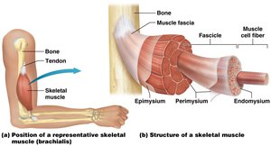
Example: The brachialis muscle in the arm demonstrates this hierarchical structure.
Fascicle Arrangement and Muscle Shapes
Types of Fascicle Arrangements
The arrangement of fascicles within a muscle affects its appearance and function. Understanding these shapes is crucial for identifying muscles and predicting their actions.
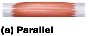
Parallel: Strap-like, evenly spaced fascicles; muscle and tendon are the same width.
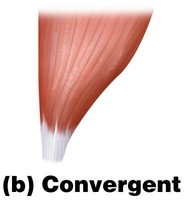
Convergent: Broad, triangular shape tapering to a single tendon.
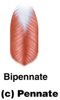
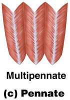
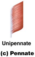
Pennate: Fascicles attach to tendon at an angle, resembling a feather.
Unipennate: Fascicles attach to one side of the tendon.
Bipennate: Fascicles attach to both sides of the tendon.
Multipennate: Several regions of fascicles joined by connective tissue.
Circular (Sphincter): Fascicles arranged in a ring, controlling body openings.
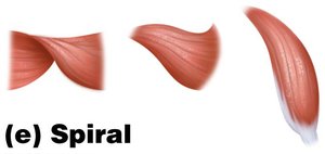
Spiral: Fascicles wrap around another structure, such as a bone.
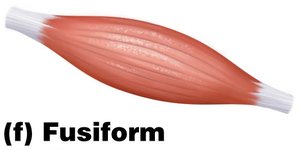
Fusiform: Muscle belly is thicker than the tapered ends.
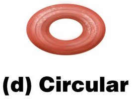
Example: The orbicularis oris is a circular muscle controlling the mouth opening.
Motor Units
Definition and Function
A motor unit consists of a single motor neuron and all the muscle fibers it innervates. The size of a motor unit determines the precision of muscle movement.
Small motor units: Few muscle fibers, allow precise movements (e.g., eye muscles).
Large motor units: Many muscle fibers, allow powerful but less precise movements (e.g., thigh muscles).
Example: Typing uses small motor units for fine finger control.
Naming Muscles
Criteria for Muscle Names
Muscles are named based on several characteristics:
Appearance: Shape or structure (e.g., deltoid = triangular).
Size: Relative size (e.g., maximus, minimus).
Location: Anatomical position (e.g., brachialis = arm).
Attachment: Bones or structures where muscles originate or insert.
Action: Function or movement produced (e.g., flexor, extensor).
Historical names: Some retain traditional names.
Example: The sternocleidomastoid is named for its attachments to the sternum, clavicle, and mastoid process.
Functions of Skeletal Muscles
Muscle Contraction and Its Roles
Skeletal muscle contractions are essential for various bodily functions:
Movement: Locomotion and manipulation of the environment.
Heat generation: Maintains body temperature (homeostasis).
Breathing: Diaphragm contraction enables respiration.
Facial expression: Muscles control expressions.
Swallowing: Muscles facilitate ingestion.
Sphincter control: Conscious regulation of body openings.
Example: Shivering increases heat production through rapid muscle contractions.
Functional Groups of Muscles
Muscle Cooperation in Movement
Muscles work in groups to produce coordinated movements:
Agonist (Prime mover): Main muscle responsible for a movement.
Antagonist: Opposes the action of the agonist, usually located on the opposite side.
Synergist: Assists the agonist by adding force or stabilizing movement.
Fixator: Stabilizes the origin of the agonist, anchoring a bone.
Example: During elbow flexion, the biceps brachii is the agonist, the triceps brachii is the antagonist, and the brachialis acts as a synergist.
Origin vs. Insertion
Anatomical Points of Muscle Attachment
Each skeletal muscle has a distinct origin and insertion:
Origin: Anchoring point, typically does not move during contraction.
Insertion: Moving end, attached to a bone or structure that moves during contraction.
Example: The origin of the biceps brachii is on the scapula, and its insertion is on the radius.
Lever Systems in Muscle Function
Types of Levers
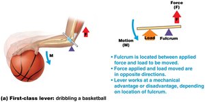
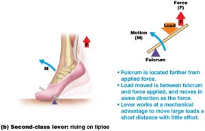
Muscles act on bones as levers, which consist of three components: load (resistance), applied force, and fulcrum (pivot point). The arrangement of these components determines the class of lever and its mechanical advantage.
First-class lever: Fulcrum between load and applied force; load moves in opposite direction to force.
Second-class lever: Fulcrum at one end, load in the middle, applied force at the other end; load moves in same direction as force.
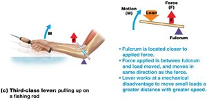
Third-class lever: Fulcrum and applied force at one end, load at the other; load moves in same direction as force.
Example: Dribbling a basketball uses a first-class lever; rising on tiptoe uses a second-class lever; pulling up on a fishing rod uses a third-class lever.
Lever Equation:
Superficial Muscles of the Body
Identification and Actions
Knowledge of the major superficial muscles, their locations, and actions is essential for understanding human movement and anatomy. Muscles are often grouped by region and function.
Head: Temporalis, Masseter
Neck: Sternocleidomastoid, Platysma
Shoulder: Deltoid
Arm: Triceps brachii, Biceps brachii, Brachialis
Forearm: Flexor carpi radialis, Extensor digitorum
Thigh: Quadriceps femoris, Sartorius
Leg: Gastrocnemius, Soleus
Abdomen: Rectus abdominis, External oblique
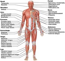
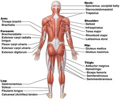
Example: The deltoid muscle abducts the arm at the shoulder.
Study Tips for Muscles
Effective Preparation Strategies
To master the muscular system, students should:
Identify major muscles using anatomical figures.
Learn muscle locations relative to bones and other landmarks.
Understand the actions of each major muscle.
Use charts and create personalized study aids for muscle actions.
Additional info: Reviewing lever systems and muscle group functions enhances understanding of movement mechanics.
