 뒤로
The Muscular System: Structure, Function, and Physiology
스터디 가이드 - 스마트 노트
 자료에 맞춘 맞춤형 노트, 핵심 정의, 예시, 맥락을 확장해 제공합니다.
The Muscular System
Overview of Muscular Tissue
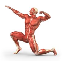
The muscular system is essential for movement, stability, and various physiological functions in the human body. Muscular tissue is classified into three main types, each with distinct structural and functional characteristics.
Skeletal Muscle: Voluntary, striated muscle attached to bones, responsible for body movements.
Cardiac Muscle: Involuntary, striated muscle found only in the heart, responsible for pumping blood.
Smooth Muscle: Involuntary, non-striated muscle found in walls of internal organs, responsible for moving substances within the body.
Microscopic Structure of Muscle Types
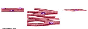
Each muscle type has unique microscopic features that relate to its function.
Skeletal Muscle: Long, cylindrical, multinucleated fibers with prominent striations.
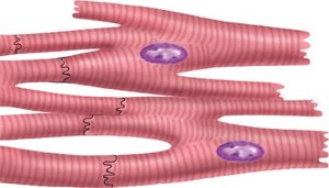
Cardiac Muscle: Branched fibers with intercalated discs, single central nucleus, and striations.
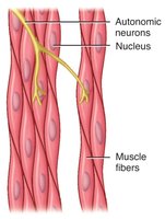
Smooth Muscle: Spindle-shaped cells with a single nucleus, lacking striations.
Characteristics of Muscular Tissue
Excitability
Muscle tissue can respond to stimuli by generating electrical signals called action potentials, similar to nervous tissue.
Excitability: Ability to respond to a stimulus and produce an action potential.
Contractility
Muscles can shorten or lengthen while generating force, enabling movement and stability.
Contractility: Ability to contract and generate force.
Extensibility
Muscle tissue can be stretched beyond its resting length without being damaged.
Extensibility: Ability to stretch or extend.
Elasticity
After being stretched or contracted, muscle tissue can return to its original shape.
Elasticity: Ability to return to original shape after stretching.
Functions of Muscular Tissue
Body Movements and Stabilization
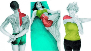
Muscles produce body movements and stabilize body positions, such as maintaining posture.
Movement: Skeletal muscles contract to move bones.
Stabilization: Muscles maintain posture and stabilize joints.
Moving Substances Within the Body
Muscles move substances such as blood, food, and waste through internal organs.
Cardiac muscle: Pumps blood.
Smooth muscle: Moves food through digestive tract, regulates blood flow.
Generating Heat (Thermogenesis)
Muscle contractions generate heat, helping maintain body temperature.
Thermogenesis: Heat production during muscle activity.
Glycemic Control
Muscles help regulate blood glucose concentration by absorbing and storing glucose.
Glycemic control: Regulation of blood sugar levels.
Anatomy of Skeletal Muscle
Organization of Skeletal Muscle
Skeletal muscle is organized into bundles of fibers surrounded by connective tissue layers.
Epimysium: Surrounds entire muscle.
Perimysium: Surrounds fascicles (bundles of fibers).
Endomysium: Surrounds individual muscle fibers.
Skeletal Muscle Fiber Anatomy
Skeletal muscle fibers are long, multinucleated cells with specialized structures for contraction.
Sarcolemma: Plasma membrane of muscle fiber.
Sarcoplasm: Cytoplasm of muscle fiber.
Sarcoplasmic reticulum: Stores calcium ions.
Myofilaments: Contractile proteins (actin and myosin).
Muscle Proteins
Types of Muscle Proteins
Muscle contraction and structure depend on three classes of proteins.
Contractile Proteins: Actin (thin filament) and myosin (thick filament) generate force.
Regulatory Proteins: Troponin and tropomyosin control contraction.
Structural Proteins: Titin, myomesin, dystrophin, actinin, and nebulin provide alignment, elasticity, and extensibility.
Regulatory Muscle Proteins
Regulatory proteins control access to myosin binding sites on actin, enabling or inhibiting contraction.
Troponin: Binds calcium ions, causing conformational change.
Tropomyosin: Covers myosin binding sites on actin when muscle is relaxed.
Excitation-Contraction Coupling
Neuromuscular Junction (NMJ)
The NMJ is the site where a motor neuron communicates with a muscle fiber to initiate contraction.
Motor neuron: Sends action potential to muscle fiber.
Acetylcholine (ACh): Neurotransmitter released into synaptic cleft.
Motor end plate: Region of sarcolemma with ACh receptors.
Excitation-Contraction Coupling Steps
Muscle contraction is triggered by electrical and chemical events at the NMJ.
Motor neuron releases ACh.
ACh binds to receptors, causing depolarization.
Action potential travels along sarcolemma.
Calcium ions released from sarcoplasmic reticulum.
Calcium binds to troponin, exposing myosin binding sites.
Myosin heads bind to actin, initiating contraction.
Muscle Cell Relaxation
Relaxation occurs when stimulation ceases and calcium is removed from the cytoplasm.
Acetylcholinesterase degrades ACh.
ATP binds myosin heads, releasing actin.
Calcium reuptake by sarcoplasmic reticulum.
Muscle Contraction Mechanisms
Sliding Filament Model and Cross-Bridge Cycling
Muscle contraction occurs as thick and thin filaments slide past each other, shortening the sarcomere.
Sliding Filament Model: Z-lines move closer, I band narrows, A band remains constant.
Cross-Bridge Cycling: Myosin heads attach, pivot, detach, and reattach to actin, powered by ATP.
Sources of Muscle Energy
ATP Production for Muscle Contraction
Muscle fibers use several mechanisms to generate ATP for contraction.
Stored ATP: Immediate energy for a few seconds.
Creatine Phosphate: Rapid ATP regeneration for short bursts.
Glycolysis: Anaerobic ATP production for moderate duration.
Aerobic Respiration: Sustained ATP production for prolonged activity.
Muscle Strength and Adaptation
Hypertrophy and Atrophy
Muscle strength is influenced by fiber size and number, which can change with use or disuse.
Hypertrophy: Increase in muscle fiber diameter due to repeated overload.
Atrophy: Decrease in fiber diameter and loss of myofibrils due to disuse or denervation.
Types of Muscle Contractions
Isotonic and Isometric Contractions
Muscle contractions can be classified based on changes in muscle length and tension.
Isotonic Contraction: Muscle changes length while tension remains constant.
Concentric Isotonic: Muscle shortens.
Eccentric Isotonic: Muscle lengthens.
Isometric Contraction: Muscle tension increases, but length does not change.
Motor Units and Control of Muscle Tension
Motor Unit Recruitment
Muscle tension is controlled by recruiting different numbers and sizes of motor units.
Motor Unit: A motor neuron and all muscle fibers it innervates.
Recruitment: Gradual activation of more motor units for increased force.
Length-Tension Relationship
The amount of stretch before contraction affects the force generated by muscle fibers.
Frequency of Stimulation
Increasing the frequency of nerve impulses can increase muscle tension and produce sustained contractions.
Muscle Tone
Definition and Clinical Significance
Muscle tone is the continuous, passive partial contraction of muscles, important for posture and joint stability.
Hypotonia: Decreased muscle tone, resulting in flaccid muscles.
Hypertonia: Increased muscle tone, resulting in rigidity.
Skeletal Muscle Fiber Types
Classification by Myoglobin Content and Function
Skeletal muscle fibers are classified based on their myoglobin content, speed of contraction, and metabolic properties.
Type | Myoglobin | Mitochondria | Glycogen | Function |
|---|---|---|---|---|
Slow Oxidative (Type I) | High | Many | Low | Endurance |
Fast Oxidative (Type IIa) | Moderate | Moderate | Moderate | Intermediate |
Fast Glycolytic (Type IIb) | Low | Few | High | Power, speed |
Exercise, Aging, and Muscle Adaptation
Effects of Exercise
Endurance and resistance exercise induce different adaptations in muscle fibers, affecting strength and fatigue resistance.
Aging and Muscle
Aging leads to a reduction in fast glycolytic fibers, muscle atrophy, and replacement of muscle tissue with connective and adipose tissue (sarcopenia).
Sarcopenia: Irreversible muscle loss, decreased quality of life, increased risk of falls.
Strength training can delay sarcopenia.
Clinical Applications
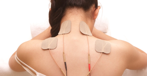
Electromyography
Electromyography measures the electrical activity of muscles to assess their function and diagnose disorders.
Neuromuscular Disorders
Botulinum toxin: Blocks ACh release, causing muscle paralysis; used medically and cosmetically.
Myasthenia gravis: Autoimmune disorder blocking ACh receptors, causing muscle weakness; treated with anticholinesterase agents.
Summary Table: Muscle Types and Characteristics
Muscle Type | Location | Control | Striations | Function |
|---|---|---|---|---|
Skeletal | Attached to bones | Voluntary | Yes | Movement, posture |
Cardiac | Heart | Involuntary | Yes | Pumping blood |
Smooth | Walls of organs | Involuntary | No | Moving substances |
