 뒤로
The Muscular System: Structure, Function, and Physiology
스터디 가이드 - 스마트 노트
 자료에 맞춘 맞춤형 노트, 핵심 정의, 예시, 맥락을 확장해 제공합니다.
The Muscular System
Overview of Muscle Tissue Types
The human body contains three main types of muscle tissue: skeletal, cardiac, and smooth muscle. Each type has distinct structural and functional characteristics that enable specialized roles in movement, posture, and internal organ function.
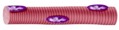
Skeletal Muscle: Voluntary, striated muscle attached to bones, responsible for body movement and posture.
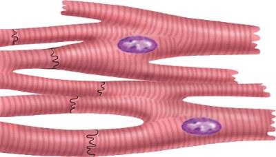
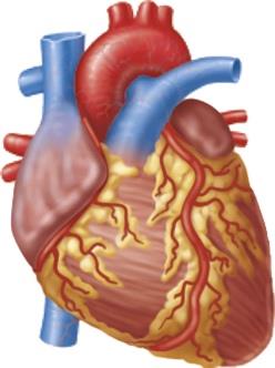
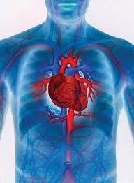
Cardiac Muscle: Involuntary, striated muscle found only in the heart, responsible for pumping blood.
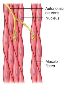
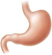
Smooth Muscle: Involuntary, non-striated muscle found in the walls of hollow organs, responsible for moving substances within the body.
Body Location and Structure of Muscle Types
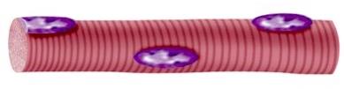
Skeletal Muscle: Attached to bones or skin; long, cylindrical, multinucleate cells with striations.
Cardiac Muscle: Located in the heart walls; branching chains of uni- or binucleate cells with striations and intercalated discs.
Smooth Muscle: Found in walls of hollow organs (e.g., stomach, intestines, blood vessels); spindle-shaped, uninucleate cells without striations.
Characteristics of Muscular Tissue
Functional Properties
Muscle tissue exhibits four key physiological properties that enable its function:
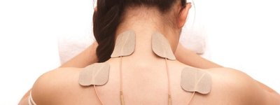
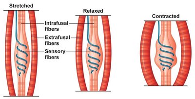
Excitability: Ability to receive and respond to stimuli by generating electrical signals (muscle action potentials).
Contractility: Ability to shorten or lengthen while generating force.
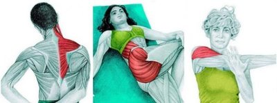
Extensibility: Ability to be stretched or extended without damage.
Elasticity: Ability to return to original shape after being stretched.
Functions of Muscular Tissue
Major Roles in the Body
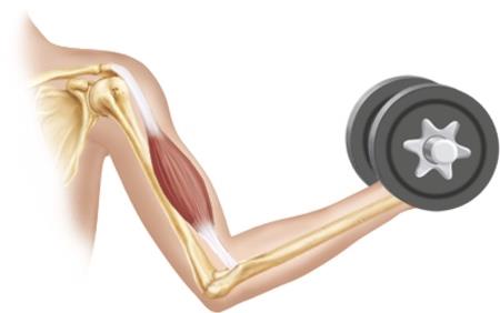
Produce Body Movements: Skeletal muscles contract to move bones and body parts.
Stabilize Body Positions & Joints: Muscles maintain posture and stabilize joints.
Move Substances Within the Body: Cardiac muscle pumps blood; smooth muscle moves food, urine, and other substances.
Generate Heat (Thermogenesis): Muscle contractions produce heat to maintain body temperature.
Glycemic Control: Muscles help regulate blood glucose by absorbing and storing glucose.


Anatomy of Skeletal Muscle
Organization and Connective Tissue Components
Skeletal muscle is organized into hierarchical structures, each surrounded by connective tissue layers:
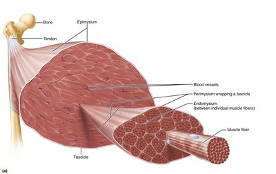
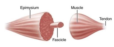
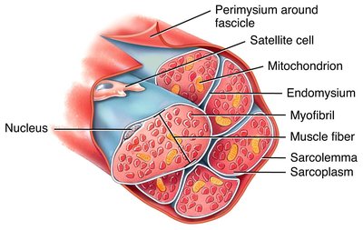
Epimysium: Surrounds the entire muscle.
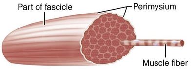
Perimysium: Surrounds bundles of muscle fibers called fascicles.
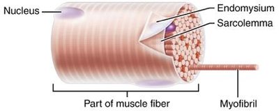
Endomysium: Surrounds individual muscle fibers (cells).
Tendon: Connects muscle to bone.
Fascia and Muscle Compartments
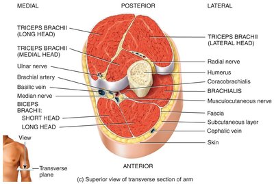
Fascia is a collagen-rich fibrous connective tissue that separates muscles or muscle groups and supports muscle compartments. It plays a crucial role in muscle organization and function.
Skeletal Muscle Fiber Anatomy
Microscopic Structure
Skeletal muscle fibers are long, multinucleated cells containing myofibrils, which are composed of repeating units called sarcomeres. Each sarcomere contains thick (myosin) and thin (actin) filaments responsible for muscle contraction.
Nerve and Blood Supply
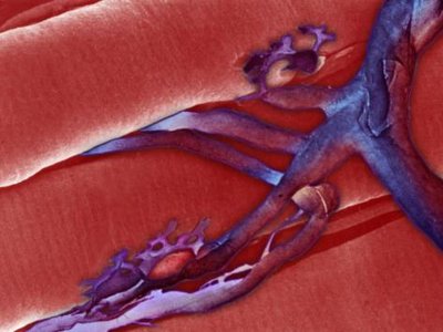
Vascularization: Muscles are well supplied with blood vessels for oxygen and nutrient delivery.
Innervation: Each muscle fiber is innervated by a motor neuron; a motor unit consists of one neuron and all the fibers it controls.
Muscle Growth and Repair
Hypertrophy: Increase in muscle fiber diameter due to repeated mechanical overload and hormonal stimulation (testosterone, growth hormone).
Hyperplasia: Increase in the number of muscle fibers; rare in humans, limited to satellite cell activity.
Atrophy: Decrease in muscle fiber diameter and loss of myofibrils due to disuse or denervation.
Fibrosis: Replacement of damaged muscle tissue with connective tissue after significant injury.

Skeletal Muscle Fiber Microscopic Anatomy
The Sarcomere and Myofilaments
The sarcomere is the basic contractile unit of muscle, defined by the area between two Z discs. It contains thick (myosin) and thin (actin) filaments arranged in a precise pattern, producing the striated appearance of skeletal muscle.
Z disc: Boundary of the sarcomere.
M line: Middle of the sarcomere, holds thick filaments together.
I band: Region with only thin filaments.
H zone: Region with only thick filaments.
A band: Length of thick filaments, including overlap with thin filaments.
Muscle Proteins
Contractile Proteins: Actin (thin filaments) and myosin (thick filaments) generate force for contraction.
Regulatory Proteins: Troponin and tropomyosin regulate contraction by controlling access to myosin binding sites on actin.
Structural Proteins: Titin, myomesin, dystrophin, α-actinin, and nebulin maintain sarcomere structure and elasticity.
Clinical Connection: Muscular Dystrophy
Muscular dystrophy is a group of genetic disorders characterized by progressive degeneration of skeletal muscle fibers due to malformation of the structural protein dystrophin, which stabilizes the sarcolemma.
Muscle Contraction Physiology
Excitation-Contraction Coupling
Excitation-contraction coupling links the electrical signal from a motor neuron to the mechanical contraction of muscle fibers. This process occurs at the neuromuscular junction (NMJ).
Action potential arrives at the axon terminal of the motor neuron.
Voltage-gated calcium channels open, triggering release of acetylcholine (ACh).
ACh binds to receptors on the sarcolemma, opening sodium channels and generating a muscle action potential.
Calcium is released from the sarcoplasmic reticulum, enabling cross-bridge cycling and contraction.
The Sliding Filament Mechanism
Muscle contraction occurs as thick and thin filaments slide past each other, shortening the sarcomere. This process is powered by ATP and regulated by calcium ions.
Cross-bridge formation: Myosin heads bind to actin.
Power stroke: Myosin heads pivot, pulling actin filaments toward the center.
Detachment: ATP binds to myosin, causing it to release actin.
Cocking: ATP hydrolysis re-energizes the myosin head.
Muscle Relaxation
Acetylcholinesterase degrades ACh, ending the signal.
Calcium is pumped back into the sarcoplasmic reticulum.
ATP binds to myosin heads, causing them to detach from actin.
Clinical Connections: Neuromuscular Disorders
Botulinum toxin: Blocks ACh release, causing paralysis.
Curare: Blocks ACh receptors, causing paralysis.
Myasthenia gravis: Autoimmune disorder blocking ACh receptors, causing muscle weakness.
Control of Muscle Tension
The Motor Unit
A motor unit consists of a motor neuron and all the muscle fibers it innervates. The size and number of motor units recruited determine the precision and strength of muscle contraction.
High precision: Few fibers per neuron (e.g., eye muscles).
Low precision: Many fibers per neuron (e.g., thigh muscles).
Twitch Contraction and Muscle Tone
Twitch contraction: Brief contraction in response to a single action potential, with latent, contraction, and relaxation phases.
Muscle tone: Continuous, partial contraction of muscles to maintain posture and readiness.
Types of Muscle Contractions
Isotonic contraction: Muscle changes length while tension remains constant.
Concentric: Muscle shortens.
Eccentric: Muscle lengthens.
Isometric contraction: Muscle tension increases, but length does not change.
Skeletal Muscle Fiber Types
Classification and Functional Differences
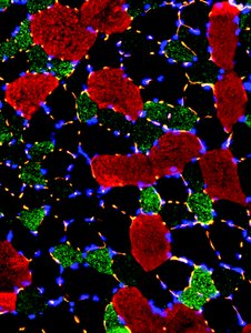
Type I (Slow Oxidative): Small, dark red, fatigue-resistant, used for endurance.
Type IIa (Fast Oxidative-Glycolytic): Intermediate size, moderately fatigue-resistant.
Type IIb (Fast Glycolytic): Large, white, powerful, fatigue quickly, used for short bursts of activity.
Muscle Response to Exercise
Aerobic exercise: Increases capillaries, mitochondria, and myoglobin; improves endurance.
Resistance exercise: Increases muscle size (hypertrophy), strength, and connective tissue.
Cardiac and Smooth Muscle Physiology
Cardiac Muscle
Contracts longer than skeletal muscle in response to a single action potential.
Relies on aerobic respiration for ATP production.
Smooth Muscle
Autorhythmic and involuntary.
Highly resistant to fatigue; maintains tone for extended periods.
Muscle Fatigue and Abnormal Contractions
Muscle Fatigue
Muscle fatigue is the inability to maintain force of contraction after prolonged activity. Causes include:
Central fatigue (CNS origin)
Peripheral fatigue (e.g., calcium depletion, lack of oxygen, nutrient depletion, lactic acid buildup)
Abnormal Contractions
Cramp: Involuntary, painful contraction due to overactive neurons or electrolyte imbalance.
Spasm: Sudden, involuntary contraction, usually painless.
Aging and Muscular Tissue
Effects of Aging
Progressive loss of skeletal muscle mass and strength.
Slower reflexes and reduced flexibility.
Relative increase in slow oxidative fibers.
Regular aerobic and strength training can slow decline.
